2Department of Acupuncture & Moxibustion, Dongguk University Bundang Oriental Hospital
3Department of Internal Korean Medicine, Dongguk University Bundang Oriental Hospital
4Department of Rehabilitation Medicine of Korean Medicine, Dongguk University Bundang Oriental Hospital
5Department of Acupuncture & Moxibustion Medicine, College of Korean Medicine, Dongguk University
Correspondence to: Won-Suk Sung, Department of Acupuncture & Moxibustion Medicine, Dongguk University Bundang Oriental Hospital 268, Buljeong-ro, Bundang-gu, Seongnam-si, Gyeonggi-do, 13601, Republic of Korea, Tel: +82-31-710-3725, E-mail: 1984sws@hanmail.net
Correspondence to: Eun-Jung Kim, Department of Acupuncture & Moxibustion Medicine, Dongguk University Bundang Oriental Hospital 268, Buljeong-ro, Bundang-gu, Seongnam-si, Gyeonggi-do, 13601, Republic of Korea, Tel: +82-31-710-3751, E-mail: hanijjung@naver.com
Received February 2, 2021 Revised February 15, 2021 Accepted February 18, 2021
The purpose of this study is to investigate the characteristics, validity, and reliability of non-radiological assessment tools of scoliosis that have been studied so far.
Methods
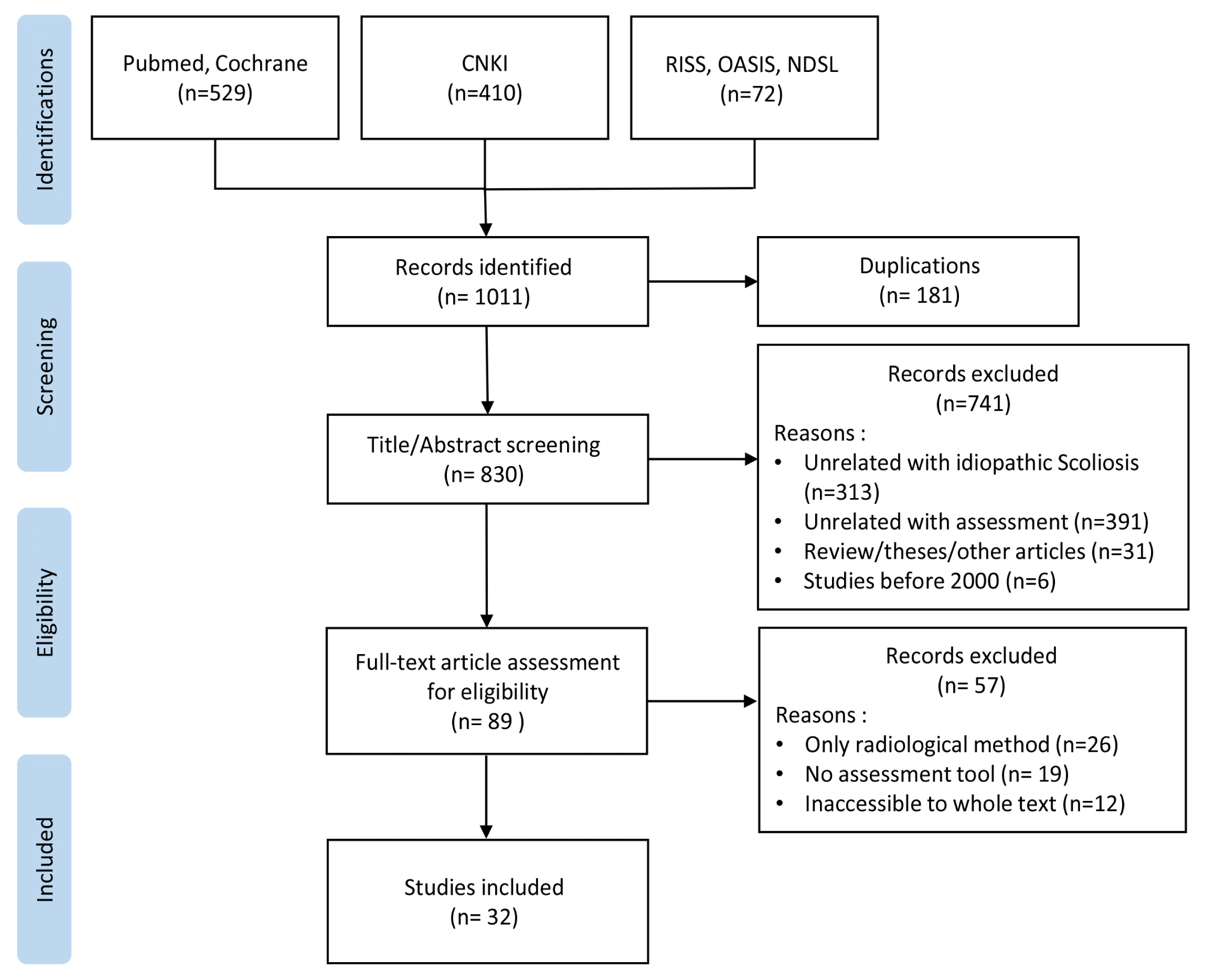
Electronic databases including Pubmed, Cochrane Library, CNKI, Science On, RISS, OASIS were searched by keywords including ‘scoliosis assessment’, ‘scoliosis screening’, ‘physical examination’, ‘functional measurement’, ‘photography’, and ‘smartphone’.
Results
32 articles using radiation-free assessments were identified from 1,011 records. The mostly used non-radiological methods were Surface topography, Scoliometer, Ultrasound, Digital Infrared Thermal Imaging, and Photography. The other methods were Gait analysis, 3D depth sensor imaging, and Low intensity electromagnetic scan.
Conclusions
It was found that non-radiological assessment tools might reduce the number of radiographs taken in scoliosis patients. To increase the reliability and validity, further research on the measurement tools of scoliosis will be needed.
3 raters on Scolioscreen-smartphone, smartphone alone and scoliometer
Franko 201234)
Scoliometer Smartphone App
Scoliometer
Cross-sectional
—
Examined 60 times with both Scoliometer and Scoliguage app
Jimbo 202035)
Hand-held roller(i-Scolioroller) combined with iPod touch
Radiography
Cross-sectional
Experiment I Plater torsos of IS patients n= 10 (1:9) age: 10 – 14 Experiment II IS patients n= 112 (15:97) age: 6 – 17
Experiment I 3 raters repeated 4 times for inter-observer reliability 8 raters measured once for intra-observer reliability Experiment II A single screening examination with i-Scrolioroller by one of 3 raters
ATI ICC of orthopedic doctor/office worker/assistant technical expert= 0.851, 0.786, 0.772 Sum ATI ICC of orthopedic doctor/office worker/assistant technical expert= 0.856, 0.900, 0.796
Prowse 201736)
Good correlation with the Scoliometer (rho= 0.78) Moderate correlation for ATR (rho= 0.627)
measured by the Baseline® Body Level/ Scoliosis meter (Examiner A ICC3,3= 1.00, Examiner B= 0.98) measured by the Scoliometer (Examiner A ICC3,3= 0.99, Examiner B= 0.98)
Correlation between CA and DHCT(difference of height cervicothoracic) r= 0.591, p<0.05 Correlation between CA and DHT(difference of height thoracic) r= 0.768, p<0.01 Correlation between CA and DHTL(difference of height thoracolumbar) r= 0.704, p<0.01 Correlation between CA and DHL(difference of height lumbar) r= 0.509, p<0.05
Cross-validation test result Accuracy of SVM to recognize scoliosis group and control group: 90.5% (if optimally selected, 95.2%) Accuracy of SVM to recognize scoliosis severity gait patterns: 81.0% (if optimally selected, 85.7%)
mean absolute difference between the paired coronal measurements: 6.3° (SD = 4.9°), Pearson’s correlation coefficient: 0.86 (P= 0.08) mean absolute difference between the paired sagittal measurements: 6.1° (SD = 4.9°), Pearson’s correlation coefficient: 0.87 (P= 0.11)
mean absolute difference between the paired coronal measurements: 2.74° (SD = 2.4°), Pearson’s correlation coefficient: 0.86 (P= 0.11) mean absolute difference between the paired sagittal measurements: 4.83° (SD = 4.28°), Pearson’s correlation coefficient: 0.87 (P= 0.67)
3-D depth sensor imaging system
Kokabu 201958)
r= 0.85 (p<0.01)
ICC Intra-class correlation coefficient, ATI Angle of trunk inclination, ATR Axial thoracic rotation
CA Cobb’s angle, DHOPI horizontal plane deformity index, POSTI posterior trunk symmetry index, PC columnar profile, RSG raster stereography, Max maximum, RMS root mean square, Rad radiography
SEM standard error of measurement, UEV upper-end vertebra, LEV lower-end vertebra, VPI-SP Volume projection imaging spinous process method, VPI-TP Volume projection imaging transverse process method
LRTA left/right trapezium angle ratio, SHA shoulder height angle, AHA axilla height angle, SVM support vector machine
Table 4
Methodological characteristics of included studies, summarizing equipments, study aims and conclusion
To evaluate the intra-and inter-observer reliability of the i-Scolioroller and to determine the optimal cutoff values of i-Scolioroller measurements
I-Scolioroller has a sufficiently high sensitivity for detecting adolescent scoliosis with a Cobb’s angle ≥20°. (Sensitivity: 88.9%, Specificity: 62.5%)
Yes
Prowse 201736)
Baseline® Body Level/Scoliosis meter (Orthopaedics Systems Incorporation®)
To investigate the reliability and validity of the Baseline® Body Level/Scoliosis meter for AIS
The Baseline® Body Level/Scoliosis meter provides reliable transverse and sagittal cervical, thoracic and lumbar measurements and valid transverse plan measurements of mild-moderate scoliosis deformity.
Yes
Sapkas 200337)
Scoliometer (Orthopaedic Systems Inc, Hayward, CA)
To create mathematic formulas that could predict the Cobb’s angle using the scoliometer measurements
Scoliometer values combined with three mathematical formulas permit assessment of adolescent idiopathic scoliosis and follow-up for progression of the deformity
Yes
Coelho 201338)
Scoliometer (Orthopaedic Systems Inc, Hayward, CA)
To measure intra- and interrater reliability, sensitivity and specificity of the scoliometer
Scoliometer and radiographic measurements showed good correlation. (The highest sensitivity value= 0.87 at 5° trunk rotation)
Digital Infrared Thermographic Imaging (T-1000 HD, MESHMEDICAL, Korea)
To investigate the correlation between Cobb’s angle and D.I.T.I on AIS
Using D.I.T.I, acupoint Simsu(BL15) is expected to be a valid indicator for the diagnosis and treatment of AIS
No
Kwok 201739)
FLIR E33 camera (FOL-18 lens; 10,800 pixels), Thermacam Researcher Professional 2.9 Software(FLIR)
To explore the possibility of using IR thermography to evaluate Infra red emissions from subjects to detect abnormalities in temperature distribution in their paraspinal muscles.
The findings of this study suggest the feasibility of incorporating IR thermography as part of SSS.
Yes
Sato 202040)
Hump measurement system with a built-in 3D camera, personal computer (Kinect for Windows: Microsoft Corporation, Redmond, Washington)
To assess the usefulness of Digital Moiré(DM) for scoliosis screening.
DM is useful as a new method for the screening of scoliosis with sufficient accuracy and reliability to replace Moiré topography. (Sensitivity= 0.98, Specificity= 0.53)
Yes
Yamamoto 201541)
—
Evaluate the accuracy of Moiré topography tool as a screening tool.
Moiré topography had a high false-positive rate (66.7%), which did not improve with examiner experience.
No
Kuroki 201842)
—
To make clear the both results and problems of SSS by Moiré topography(MT)
SSS by MT seemed to be effective in detecting scoliosis although both positive predictive value and the reference rate to the second screening were low.
Yes
Choi 200543)
3D-surface topography (Koastron, IBS-2000, Korea)
To measure correlation between 3D-surface topography and Cobb’s angle in scoliosis
3D surface topography and Cobb’s angle was highly correlated.
To assess the usefulness of Surface topography(ST) for scoliosis screening
Did not reveal the advantage of ST as a scoliosis screening method in comparison with the use of scoliometer.
No
Komeili 201444)
Four VIVID 910 3D laser scanners (Konica Minolta Sensing Inc., Ramsey, NJ, USA), Polygon Editing Tool (PET version 2.21, Konica Minolta), Geomagic software (Geomagic Studio 12, Morrisville, NC, USA)
Introduces a 3D markerless analysis technique for assessing torso asymmetry in AIS and a system for classifying patients based on this technique.
Distinctive patterns of asymmetry were identified with very good to excellent reliability.
Yes
Pino-Almero 201745)
Mobile white screen, projector, digital camera, computer with image recognition software designed in Matlab 7.9.0 (Matlab & Simulink Release 2009b. The Mathworks, Inc., Natick, MA, USA)
To study the correlation between asymmetry of back (measured by ST) and deformity of the spine (quantified by Cobb’s angle)
ST cannot substitute for radiographs in the diagnosis of scoliosis but it can offer data that complement radiologic study.
Mobile white screen, projector EPSON(3LCD projector model: EMP-835), digital camera CANON, computer(MacBook Pro) with the program developed in MATLAB 7.9.0
To study if ST would be useful in the follow-up of AIS patients
ST showed 90.32% agreement with radiographic method in follow-up of AIS patients.
Yes
L.Schulte 200846)
Formetric system (Diers International, Wiesbaden, Germany)
To investigate the reliability and accuracy of raster stereography in comparison with radiography as the gold standard.
Rasterstereography accurately reflects the radiographically measured progression of scoliosis during the long-term follow-up, but these parameters are not directly comparable with the Cobb’s angle.
Yes
Drzal-Grabiec 201447)
Mora projection (MORA System 4th Generation)
To evaluate the compatibility of external measurements of parameters characterizing scoliosis using the photogrammetric method.
The photogrammetric method gives significant results in terms of parameters characterizing the position of the shoulder blades and shoulders, as well as pelvis rotation.
To evaluate validity and reliability of Ultrasound imaging compare to MRI
The ultrasound imaging is a reliable and valid measurement of spinal curvature in the coronal plane using Center of Laminae (COL) method.
Yes
Cheung 201549)
Ultrasound scanner EUB-8500 (Hitachi Ltd., Tokyo, Japan), a computer with an Intel Core 2 Q6600 2.4-GHz processor and a video capture card NIIMAQPCI/PXI-1411 (National Instruments Corporation, Austin, TX, USA), a compact electromagnetic spatial sensing device MiniBird Model 130 (Ascension Technology Corporation, Burlington, VT, USA).
To assess the performance of newly developed freehand 3D ultrasound system.
Results suggested that the ultrasound volume projection imaging method can be a promising approach for the assessment of scoliosis.
Yes
Jiang 201950)
A custom-designed liner 2-D ultrasound probe (width: 10 cm; frequency: 4–10 MHz), An electromagnetic spatial sensing device (driveBAY, Ascension Technology Corporation, Burlington, USA)
To develop a fast 3-D ultrasound projection imaging (FUPI) method for assessment of scoliosis.
The results indicate that the developed projection imaging method could greatly decrease the processing time while preserving the comparative image quality.
Yes
Zheng 201651)
The Scolioscan system (Model SCN801, Telefield Medical Imaging Ltd, Hong Kong)
To test the reliability of spine deformity measurement of Scolioscan and its validity compared to the Cobb’s angle from radiography in AIS patients.
Scolioscan is reliable for measuring coronal deformity for patients with AIS and appears promising in screening large numbers of patients, for progress monitoring, and evaluation of treatment outcomes.
To demonstrate the reliability of using diagnostic ultrasound imaging(USI) in the assessment of the thickness of the soft tissues of the interscapular region in AIS
USI could be a reliable method in evaluating of the thickness of the soft tissues of the interscapular region which in turn could be a useful guide to the assessment of the effects of AIS.
Yes
Matmalas 201452)
Digital Nikon D5100 (Nikon Corporation, Tokyo, Japan) camera
To determine the validity of digital photography as an evaluation method for shoulder balance (ShB) in patients with idiopathic scoliosis.
Digital clinical photography appears to be a reliable method for objective clinical measurement of ShB. The correlation between clinical and radiological balance is statistically significant although moderate/weak.
No
Aroeira 201153)
A digital camera, Sony 7.1 megapixel (Sony, Manaus, Amazonas, Brazil), a Greika WT3750 (Greika, São Paulo, SP, Brazil) tripod, A Carci Simetograph (Carci, Americanópolis, SP, Brazil), CorelDraw13 software (CorelCorporation, Ottawa, Canada)
To develop a protocol for computerized photogrammetry for the quantification of scoliosis, and to mathematically relate this proposed method with the Cobb radiographic method.
The preliminary results presented demonstrate equivalence between the two methods. More studies are needed.
Yes
Saad 200954)
Photographic camera positioning (Sony P200 7.2.mp; Sony, Tokyo, Japan)
The purpose of this study was to investigate the reliability and validity of photogrammetry in measuring the lateral spinal inclination angles.
Although the current study did not show the validity of photogrammetry as a measure of the lateral spinal curvature in scoliosis, high reliability coefficients were observed.
No
Kim 201455)
The Smartstep™ pneumatic insole, The Smartstep™ software
To demonstrate that relationship between scoliosis and gait factor and foot weight bearing in ambulation.
In this study Influence of scoliosis was not found on the rate of stance phase and rate of swing phase and gait velocity. Fore foot weight bearing (P = 0.019) was significantly higher in the AIS group.
No
Cho 201856)
IMU-based system (Human Track, Rbiotech Co., Ltd., Seoul, Korea) consisting of a gyroscope, accelerometer and magnetic sensor
This study discussed application of a machine learning approach for the automatic cognition of gait changes due to scoliosis using gait measures.
Study’s results demonstrate considerable potential in applying SVMs in gait classification for medical applications. (Accuracy of SVM to recognize scoliosis group and control group: 90.5% ) (Accuracy of SVM to recognize scoliosis severity gait patterns: 81.0% )
Yes
Ovadia 200757)
Ortelius800™ system (OrthoScan Technologies, Rosh Pina, Israel)
To investigate the clinical value of Ortelius800™
Found the novel clinical tool to be reliable for following mild and moderate idiopathic curves in both coronal and sagittal planes.
Yes
Kokabu 201958)
Consumer-grade 3D depth sensor (Xtion Pro Live, ASUSTeK Computer Inc. Taipei, Republic of China), a laptop computer (Core-i5, 7200U-4 GB HP pavilion-15-au105tu, HP Inc, California, USA)
To report the potential accuracy of newly developed, asymmetry-recognition system for the surface of the human back using a 3D depth sensor
This study demonstrates the outstanding ability of this new system for deciding whether additional radiography is needed to define scoliosis. This system can be an alternative to the forward bend test and scoliometer measurement in clinics. (Sensitivity: 0.97, Specificity: 0.93)
Yes
D.I.T.I digital infrared thermographic Image, IR infrared, SSS school scoliosis screening, AIS adolescent idiopathic scoliosis
ST surface topography, AIS adolescent idiopathic scoliosis
SVM support vector machine
참고문헌
1. Hresko MT. Idiopathic scoliosis in adolescents. New England Journal of Medicine. 2013; 368:9. 834–41.
2. Bunnell WP. The natural history of idiopathic scoliosis before skeletal maturity. Spine. 1986; 11:8. 773–6.
3. Cobb J. Outline for the study of scoliosis. Instr Course Lect AAOS. 1948; 5:261–75.
4. Doody MM, Lonstein JE, Stovall M, Hacker DG, Luckyanov N, Land CE. Breast cancer mortality after diagnostic radiography: findings from the U.S. Scoliosis Cohort Study. Spine (Phila Pa 1976). 2000; 25:16. 2052–63.
5. Goldberg MS, Mayo NE, Levy AR, Scott SC, Poîtras B. Adverse reproductive outcomes among women exposed to low levels of ionizing radiation from diagnostic radiography for adolescent idiopathic scoliosis. Epidemiology. 1998; 271–8.
6. Schmitz-Feuerhake I, Pflugbeil S. ‘Lifestyle’ and cancer rates in former East and West Germany: the possible contribution of diagnostic radiation exposures. Radiation protection dosimetry. 2011; 147:1–2. 310–3.
7. Legaye J. Follow-up of the sagittal spine by optical technique. Annals of physical and rehabilitation medicine. 2012; 55:2. 76–92.
8. Mínguez MF, Buendía M, Cibrián RM, Salvador R, Laguía M, Martín A, et al. Quantifier variables of the back surface deformity obtained with a noninvasive structured light method: evaluation of their usefulness in idiopathic scoliosis diagnosis. European Spine Journal. 2007; 16:1. 73–82.
9. Pruijs J, Hageman M, Keessen W, Van Der Meer R, Van Wieringen J. Variation in Cobb angle measurements in scoliosis. Skeletal radiology. 1994; 23:7. 517–20.
10. Carman D, Browne R, Birch J. Measurement of scoliosis and kyphosis radiographs. Intraobserver and interobserver variation. The Journal of bone and joint surgery. American volume. 1990; 72:3. 328–33.
11. Jae-Ho Choi G-BK, Kim Sang-Hyun, Kim Gyoo-Hyung, Lee Mi-Hwa, Ahn Jung-Seong, Hong Seong-wan, Lee Jae-Seok, Kwon Ick-Su. The Study on the Perceptions of Radiological Technologist in Medical Imaging Equipment Used by the Oriental Doctor. 2017; 40:1. 109–20.
12. Bunnell WP. An objective criterion for scoliosis screening. J Bone Joint Surg Am. 1984; 66:9. 1381–7.
13. Dangerfield P, Denton J, Barnes S, Drake N. In : The assessment of rib-cage and spinal deformity in scoliosis in Proceedings of the 4th International Symposium on Moiré Fringe Topography and Spinal Deformity; Oxford Gustav Fischer Verlag. 1987.
14. Amendt LE, Ause-Ellias KL, Eybers JL, Wadsworth CT, Nielsen DH, Weinstein SL. Validity and reliability testing of the Scoliometer. Phys Ther. 1990; 70:2. 108–17.
15. Upadhyay SS, Burwell RG, Webb JK. Hump changes on forward flexion of the lumbar spine in patients with idiopathic scoliosis. A study using ISIS and the Scoliometer in two standard positions. Spine (Phila Pa 1976). 1988; 13:2. 146–51.
16. Murrell GA, Coonrad RW, Moorman CT, Fitch RD. An assessment of the reliability of the Scoliometer. Spine (Phila Pa 1976). 1993; 18:6. 709–12.
17. Grivas TB, Vasiliadis ES, Mihas C, Triantafyllopoulos G, Kaspiris A. Trunk asymmetry in juveniles. Scoliosis. 2008; 3:13
18. Kotwicki T, Chowańska J, Kinel E, Lorkowska M, Stryła W, Szulc A. Sitting forward bending position versus standing position for studying the back shape in scoliotic children. Scoliosis. 2007; 2:1. S34
19. Choi YC CL, Kwon KR. Standardization Study of Thermal Imaging using the Acupoints in Human Body. Journal of pharmacopuncture. 2008; 11:3. 113–22.
20. Haddad DS, Brioschi ML, Arita ES. Thermographic and clinical correlation of myofascial trigger points in the masticatory muscles. Dentomaxillofac Radiol. 2012; 41:8. 621–9.
21. Yang TJ, Jeong SJ, Kwak MK, Jang YJ, Hyun MK, Yoon TK, et al. A Clinical Study on Adolescent Idiopathic Scoliosis using DITI. The Acupuncture. 2016; 33:4. 7–14.
22. Kwon GR KH. The standardization study for the oriental clinical application of infrared body-heat measurement image I. The Acupuncture. 1996; 13:2. 1–22.
23. Bae Eun-jung SJ-c, Sung-chul Lim, Sang-won Han. A Clinical Study on Diahnosis of the patients with Scoliosis by D.I.T.I. The Journal of Korean Acupuncture & Moxibustion Society. 2004; 21:1. 51–8.
24. Kim JM. Clinical Application of Infrared Thermography. The Society of Korean Medicine Diagnosis. 2000; 4:1. 32–42.
25. Weisz I, Jefferson RJ, Turner-Smith AR, Houghton GR, Harris JD. ISIS scanning: a useful assessment technique in the management of scoliosis. Spine (Phila Pa 1976). 1988; 13:4. 405–8.
26. Knott P, Mardjetko S, Nance D, Dunn M. Electromagnetic topographical technique of curve evaluation for adolescent idiopathic scoliosis. Spine (Phila Pa 1976). 2006; 31:24. E911–5. discussion E6.
27. Chowanska J, Kotwicki T, Rosadzinski K, Sliwinski Z. School screening for scoliosis: can surface topography replace examination with scoliometer? Scoliosis. 2012; 7:1. 9
28. Pino-Almero L, Mínguez-Rey MF, Sentamans-Segarra S, Salvador-Palmer MR, de Anda RMC-O. Quantification of topographic changes in the surface of back of young patients monitored for idiopathic scoliosis: correlation with radiographic variables. Journal of biomedical optics. 2016; 21:11. 116001
29. Factor D, Perlas A. Ultrasound-assisted lumbar plexus block in a patient with scoliosis. Reg Anesth Pain Med. 2010; 35:6. 568–9.
30. Hyo-Jeong K, Wan-Hee KS-YL. Reliability of Ultrasound Imaging of the Thickness of the Soft Tissues of the Interscapular Region in Adolescent Idiopathic Scoliosis. Special Education & Rahabilitation Science Research Center. 2012; 51:4. 177–88.
31. Zhang J, Li H, Yu B. Correlation between Cobb Angle and Spinous Process Angle Measured from Ultrasound Data. In : Proceedings of the 2017 4th International Conference on Biomedical and Bioinformatics Engineering; 2017.
32. Thomsen M, Abel R. Imaging in scoliosis from the orthopaedic surgeon’s point of view. Eur J Radiol. 2006; 58:1. 41–7.
33. Cohen L, Kobayashi S, Simic M, Dennis S, Refshauge K, Pappas E. Non-radiographic methods of measuring global sagittal balance: a systematic review. Scoliosis Spinal Disord. 2017; 12:30