Due to advance of science and IT technology, high tech imaging equipment like ultrasonography, CT, MRI and PET scan is constantly being developed and progressed; use of these techniques is needed for Korean medicine.
Methods:
Historical study was undertaken on the evidence of using ultrasonography. Normal organs and various sorts of diseases were also observed by ultrasonography.
Results:
Korean medicine judges disease of internal organs and condition of health by symptoms of functional disease and organic disease based on anatomical theory. Ultrasonography based on anatomical theory is non-invasive and free from radiation exposure and can be performed directly from clinical practice in real-time. Ultrasonography can be a big help for securing the stability of the internal organs in inserting needles in the thorax and abdomen as well as diagnosing functional and organic diseases based on anatomical theory.
Conclusion:
We look forward to a great development of scientification and objectification of Korean medicine by using and researching imaging equipment based on anatomical theory as well as ultrasonography.
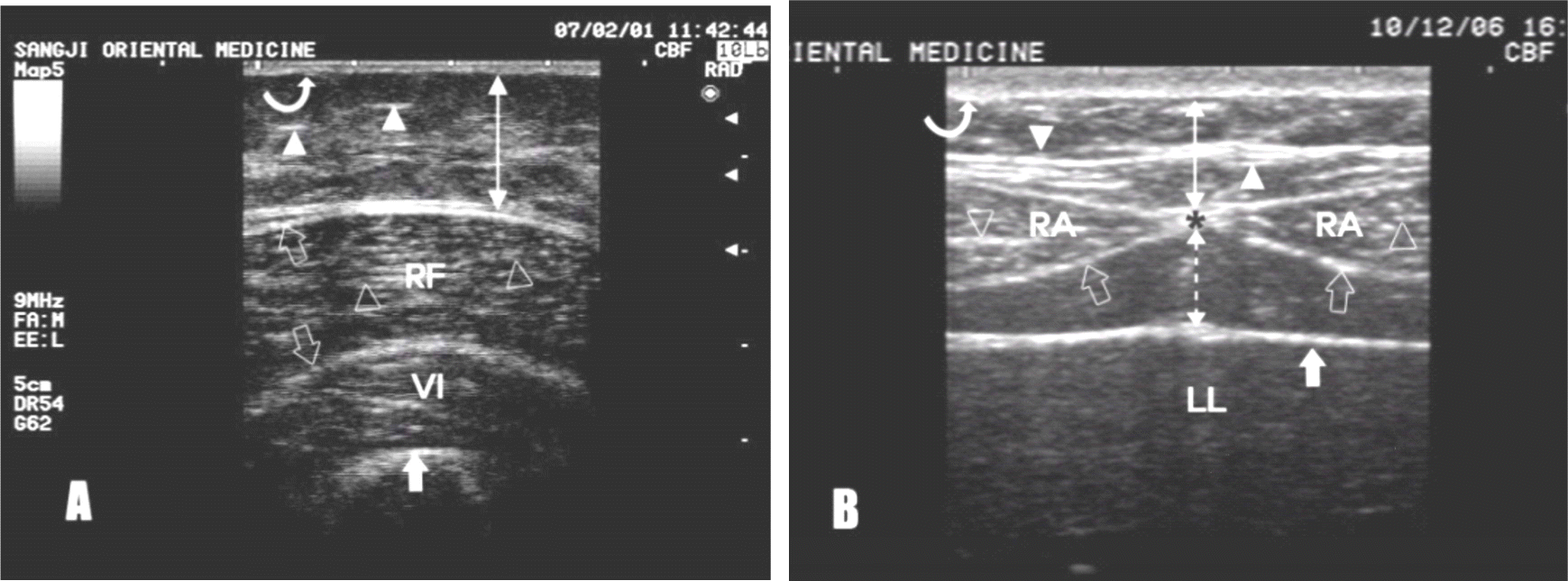
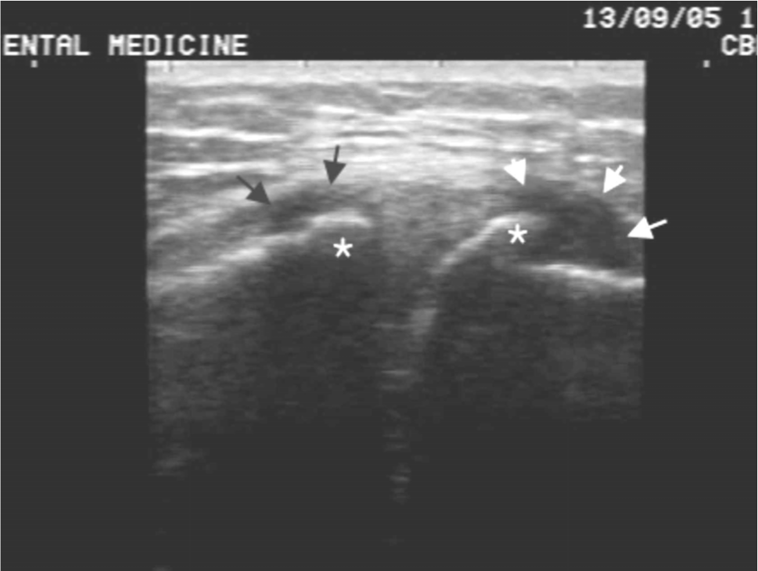
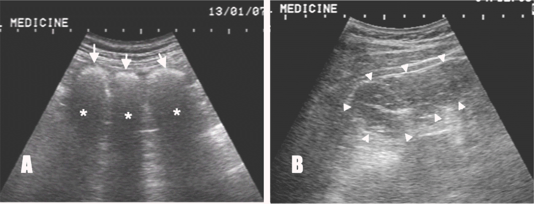
Transverse ultrasonography findings of normal thigh(A) and abdominal wall(B) curved arrow: skin(epidermis and dermis), double arrow: fat of subcutaneous layer, dotted double arrow: preperitoneal fat, white arrow heads: connective tissue septa, open arrows: muscle fascia, open arrow head: fibroadipose septa, white arrow: surface(A) of femur bone and peritoneum(B), black asterisk: linea alba, RF: rectus femoralis muscle, RA: rectus abdominalis, VI: vastus intermedius muscle, LL: left lobe of liver
Fig. 2.
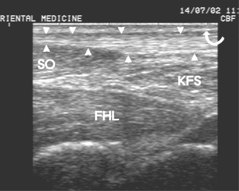
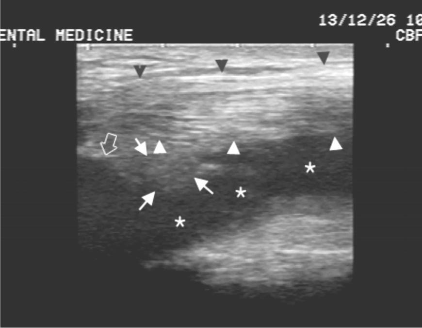
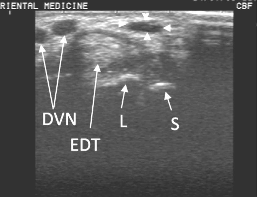
Longitudinal ultrasonography finding of normal geun(筋) curved arrow: skin(epidermis and dermis), arrow heads: achilles tendon, SO: soleus muscle, FHL: flexor hallucis longus muscle, KFS: Kager fat space
Fig. 3.
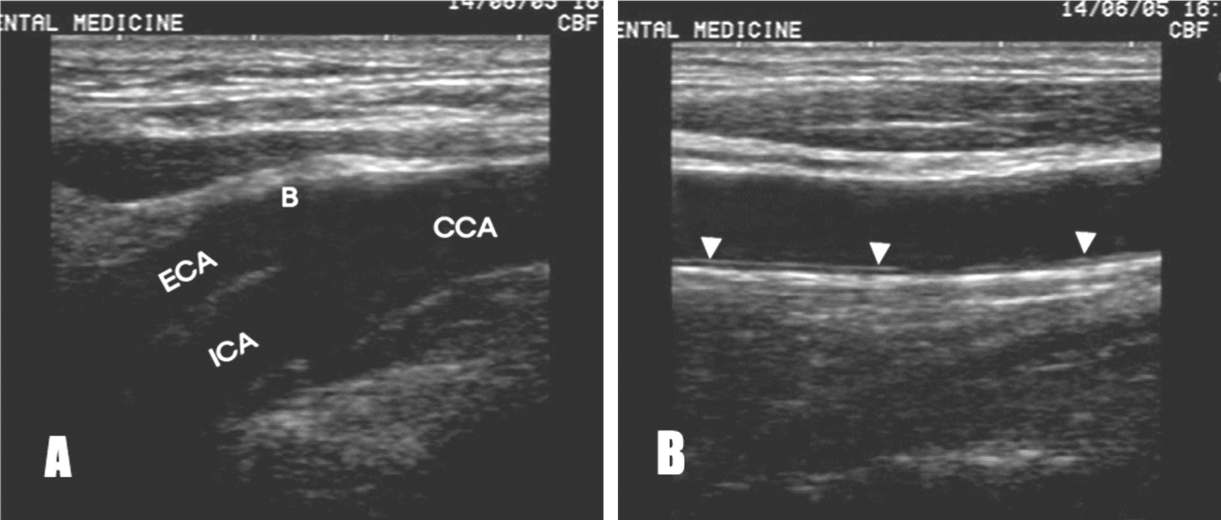
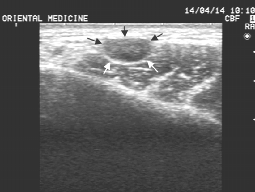
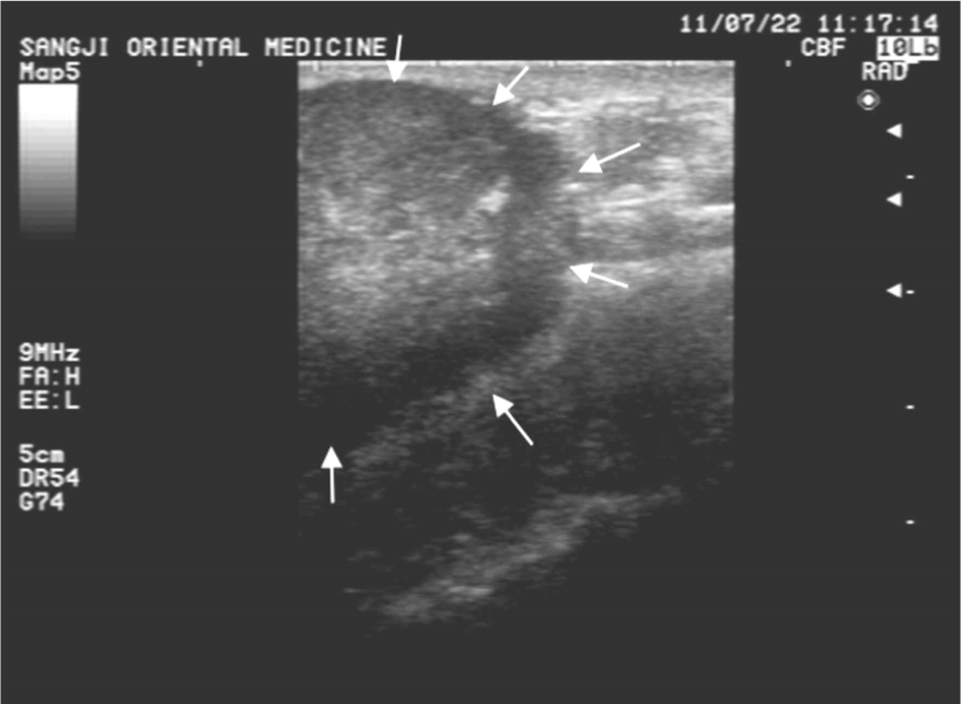
Longitudinal ultrasonography findings of normal common carotid bifurcation(A) and wall of CCA(B) around inyeong-maek(人迎脈)
Ultrasonography finding of IMT of inyeong-maek(人迎脈)
US image of IMT of the CCA shows normal range within 1mm
US: ultrasonography, IMT: intima-media thickness,
CCA: common carotid artery
Fig. 5.
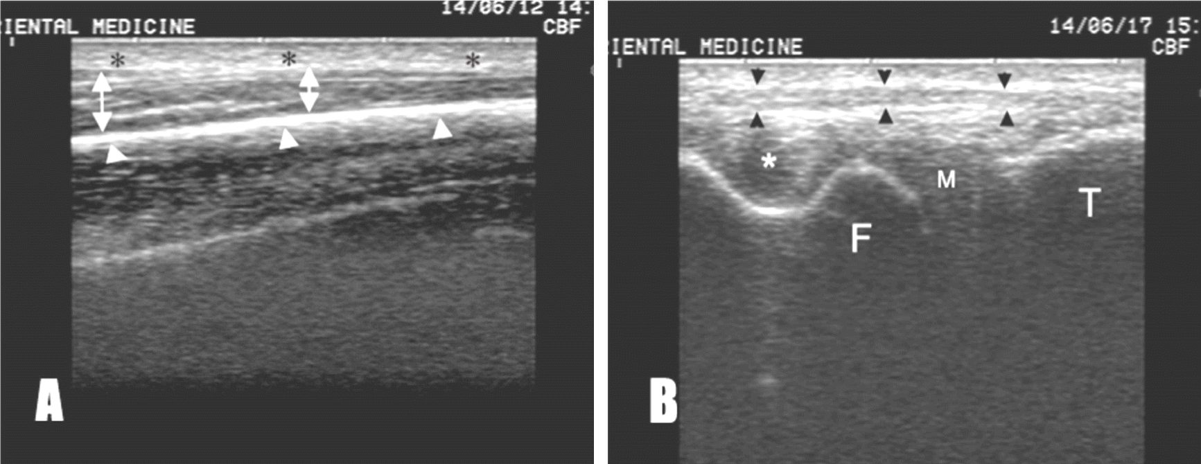
Ultrasonography findings of ulna bone(A) and lateral knee joint of knee (B) black asterisks: skin, double arrow: subcutaneous layer, white arrow heads: surface of ulnar bone, black arrow heads: lateral collateral ligament, white asterisks: popliteus tendon, M: miniscus, F: femor bone, T: tibia bone
Fig. 6.
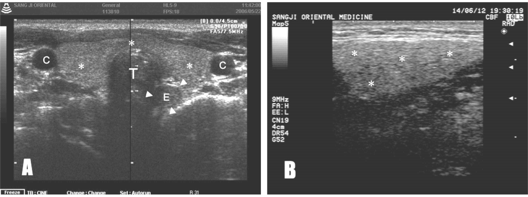
Ultrasonography findings of thyroid glands(A) and submandibular salivery gland(B) white asterisks: thyroid, double asterisks: submandibular gland, E:esophagus, arrow heads: esophagus wall
Fig. 7.
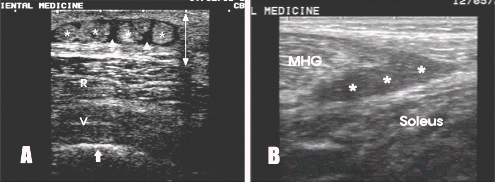
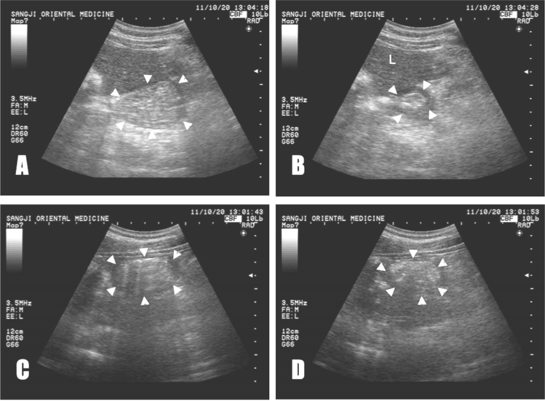
Ultrasonography findings of eohyeol(瘀血)
(A) Transverse US image of the thigh shows hematoma(arrow head) with detached fat fragments(asterisks).
(B) Longitudinal US image of the medial head of gastrocnemius muscle shows hypoechoic lesion(asterisks) with hemorrhagic infiltration, white arrow: surface of femur bone, R: rectus femoralis muscle, V: vastus intermedius muscle
Fig. 8.
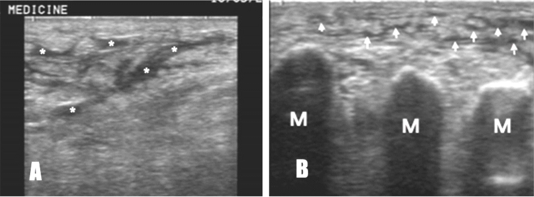
Ultrasonography findings of bujong(浮腫) of the shin(A) and the dorsum of foot(B)
(A) US image of the shin shows the fluid collections of the lymphatic channel(asterisks). (B) US image of the dorsum of foot shows the fluid collections of the lymphatic channel(arrows).
M: metatarsus
Fig. 9.
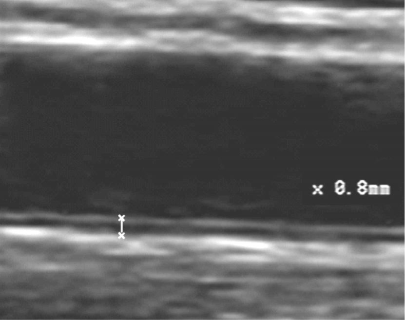
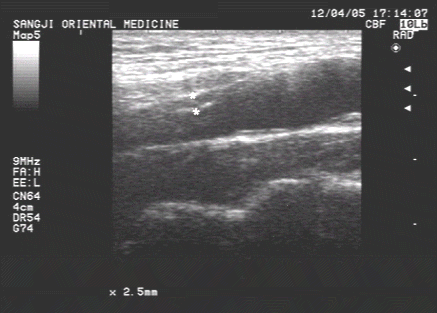
Ultrasonography finding of increased IMT of inyeong-maek(人迎脈)
US image of the internal carotid artery shows the increase of the IMT with 2.5mm.
IMT: intima-media thickness
Fig. 10.
Ultrasonography findings of stenosis(A) and plague(B) of inyeong-maek(人迎脈)
(A) Transverse US image of the CCA shows severe stenosis with hypoechoic atheroma(arrows).
(B) Longitudinal US image of the CCA shows plague with hyperechoic plague(arrows).
Fig. 11.
Transverse(A) and longitudinal(B) ultrasonography findings of bujong(浮腫) around geun(筋) US image shows effusion of biceps tendon sheath.
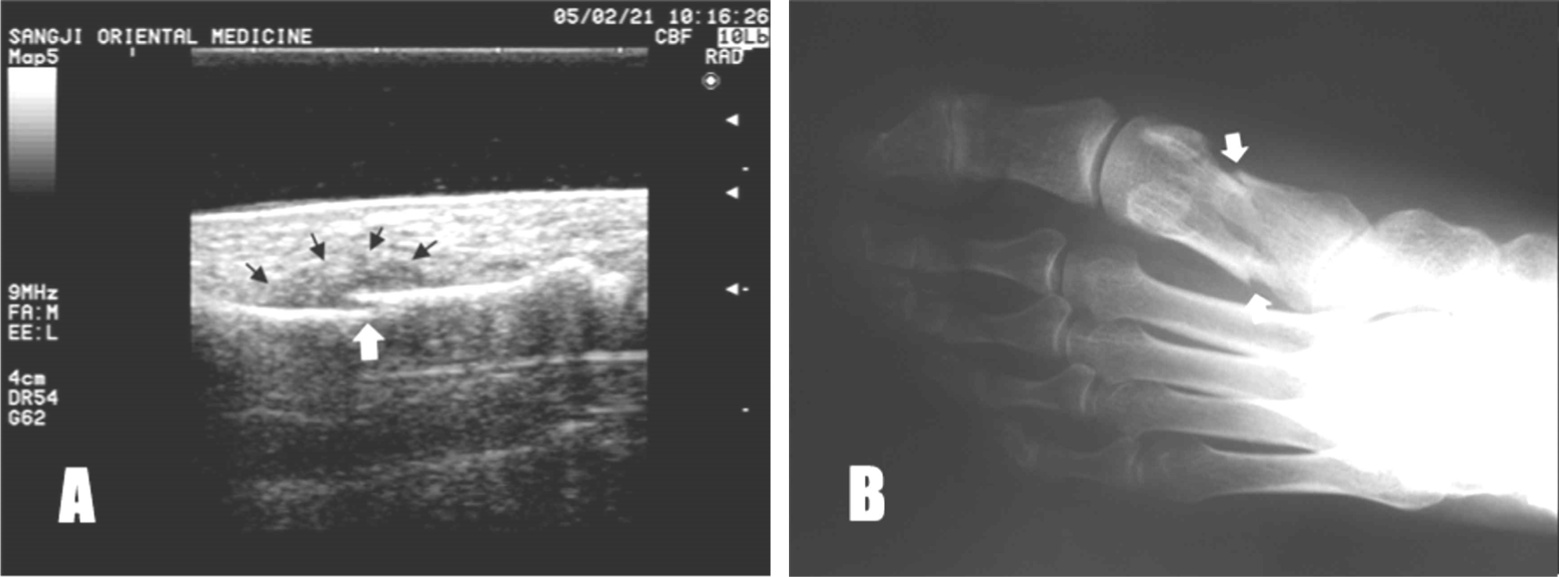
Ultrasonography and X-ray findings of goljeol(
骨折) of 1st metatarsal bone
(a) US image demonstrates focal breaks of hyperechoic cortical line(white arrow) of the 1st metatarsal bone surface with overlying soft tissue edema(black arrows), (b) X-ray image demonstrates fracture of the 1st metatarsal bone(white arrows).
Fig. 13.
Ultrasonography finding of hakseulpung(鶴膝風) Longitudinal US image of medial knee shows osteophytes(asterisks) and joint effusion(arrows).
Fig. 14.
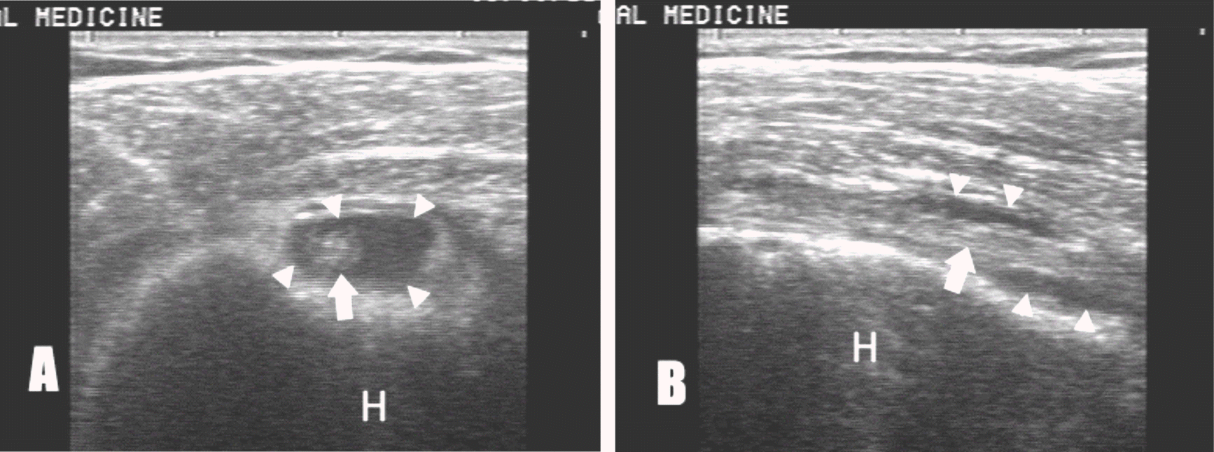
Ultrasonography finding of yeokjeolpung(歷節風)
Longitudinal US image of anterior knee shows patella(open arrow), fat pad(arrows), quadriceps tendon(arrow heads) and suprapatellar bursa effusion(asterisks).
Fig. 15.
Ultrasonography findings of yeongbyeong(癭病)
(A) US image shows enlarged thyroid gland(asterisks). (B) US image shows hypoechoic mass(arrow heads) in thyroid gland, which turned out to be malignant.
C: common carotid artery, T: trachea
Fig. 16.
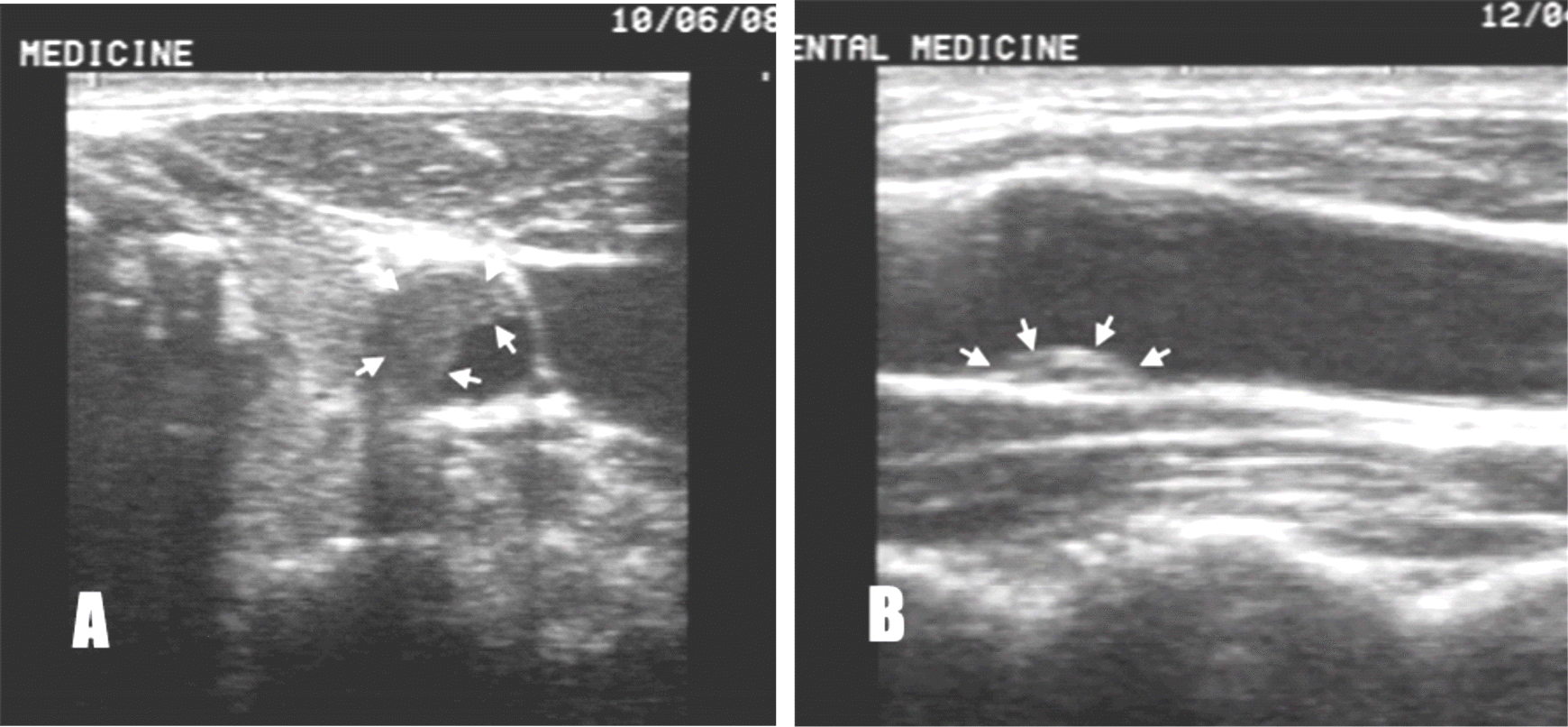
Ultrasonography finding of naryeok(瘰癧)
US image shows benign lymph node enlargement(arrows) about 12mm with an regular hypoechoic oval-shape at the neck.
Fig. 17.
Ultrasonography finding of damhaeg(痰核) US image shows ganglion(arrow heads) about 6mm with an regular anechoic oval-shape at the wrist.
US image shows heterogeneous hypoechoic huge mass(arrows), which turned out to be breast cancer.
Fig. 19.
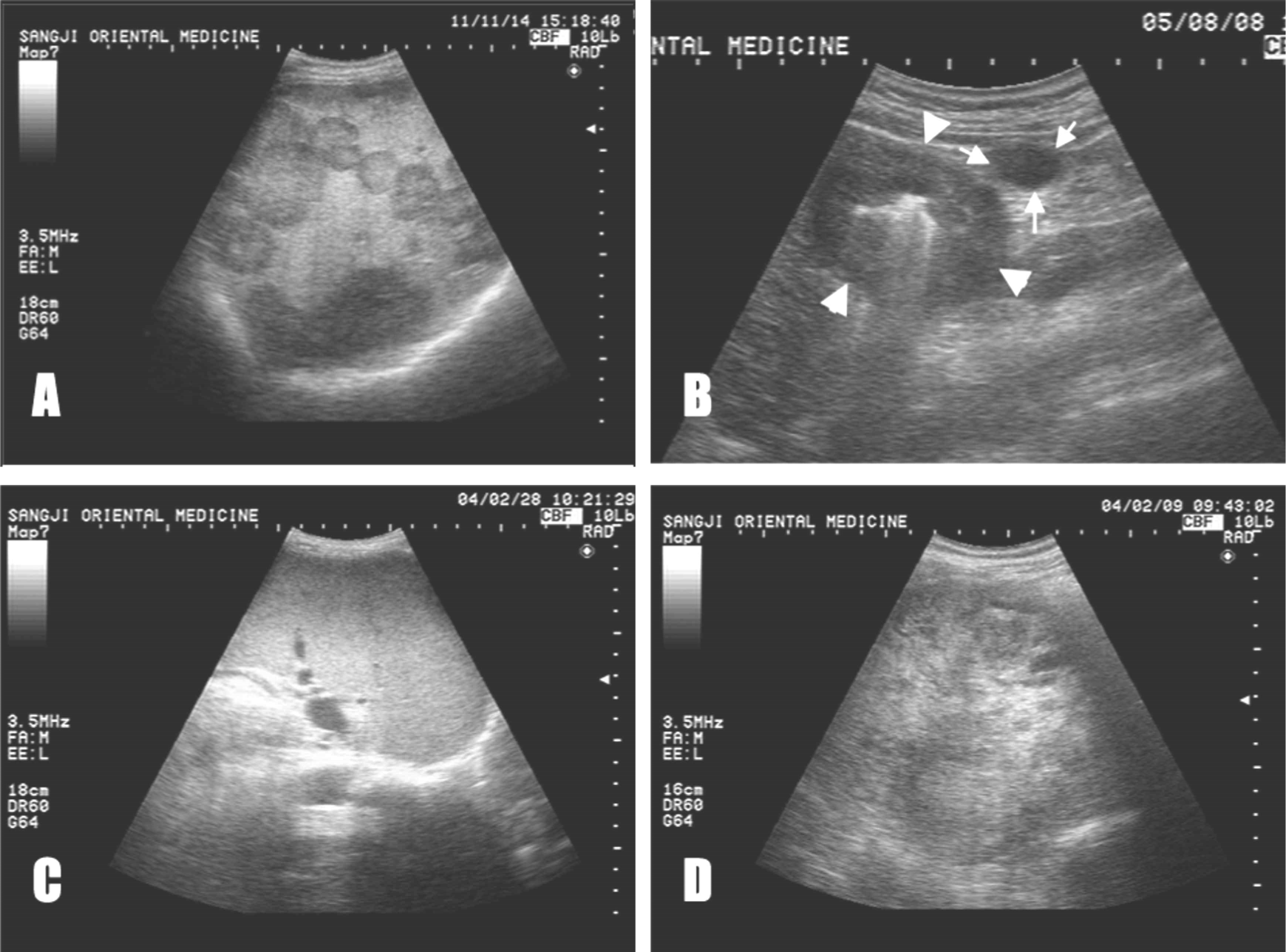
Ultrasonography findings of jeokchwi(積聚)
(A) Metastatic liver cancer with hepatomegaly (B) Longitudinal image of the pyrolus of stomach shows hypoechoic wall thickening(pseudokedney sign,arrow heads) due to gastric cancer with lymphadenopathy(arrows). (C) Splenomegaly (D) Huge kidney cancer
Fig. 20.
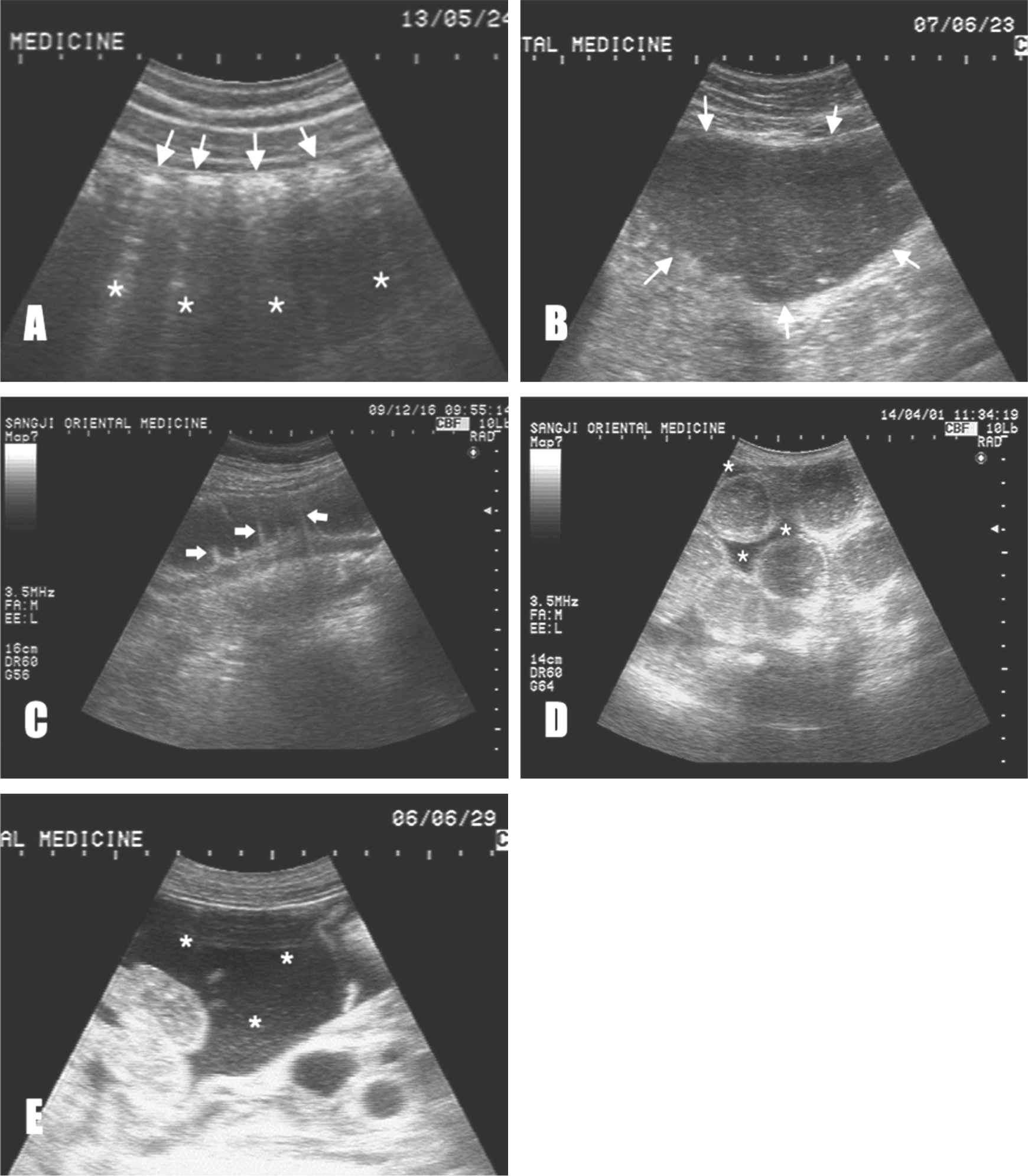
Ultrasonography findings of changman(腸滿)
(A) normal colon gas pattern : US image shows hyperechoic short lines(arrows)due to gases and dirty shadow(asterisks) secondary to reflection. (B) severe gastric distention (arrows) (C) dilatation of jejunum due to ileus with prominent valvulae conniventes(arrows) (D) dilatation of ileum due to ileus with small amounts of ascites(asterisks) (E) massive acites (asterisks) due to liver cirrhosis
Fig. 21.
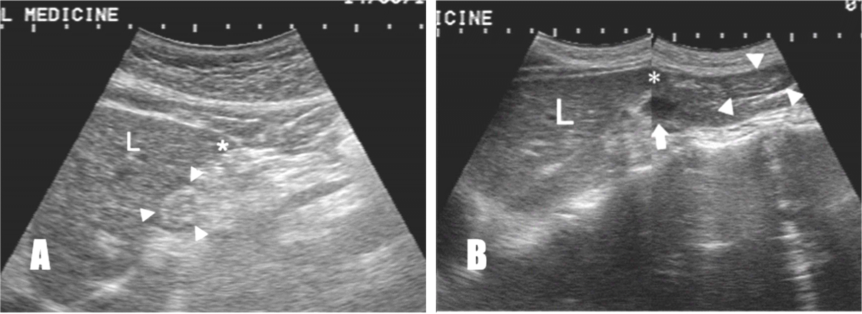
Longitudinal ultrasonography findings of normal person(A) and wiha(胃下) person(B)
(A) US image shows normal pylorus(arrow heads)which is above from inferior border angle of left lobe of live(asterisk). (B) US image shows drooped pylorus(arrow heads)which is below from inferior border angle of left lobe of live(asterisk).
L: left lobe of live, white arrow: splenic vein
Fig. 22.
Longitudinal ultrasonography findings of pyloric part of stomach about 10 minutes after meal on normal person(A,B) and wiwan(胃緩) person(C,D)
(A) pylorus extension (B) full pylorus contraction about 10 seconds after pylorus extension (C) pylorus extension (D) incomplete pylorus contraction about 10 seconds after pylorus extension
Fig. 23.
Ultrasonography findings of constipation(便秘)(A) and diarrhea(泄瀉) (B) in the colon
(A): US image shows hyperechoic arc lines(arrows) due to gases and clean acoustic shadows(asterisks) secondary to reflection due to hard stool. (B) US image shows anechoic fluid collection in the colon(arrow heads).
Fig. 24.
Ultrasonography findings of bladder of normal person(A) and yobulri(尿不利) person(B)
(A) US image show normal bladder(asterisks) and prostate. (B) US image show bladder wall thickening(arrow heads) due to chronic cystitis with BPH.
P: prostate, BPH: benign prostatic hyperplasia
Fig. 25.
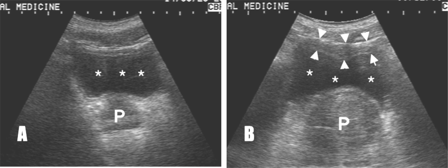
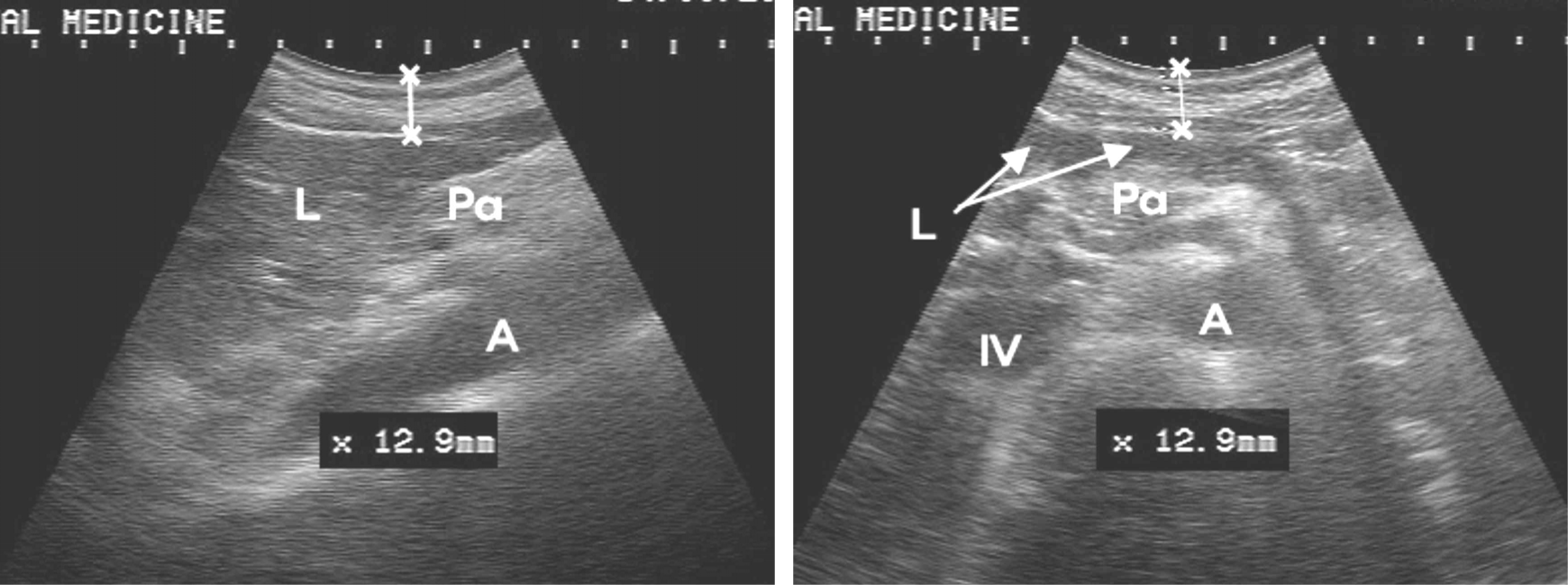
Ultrasonography findings on jungwan-hyeol(CV12,
中脘穴) of 42kg–150cm woman
(a) Longitudinal US image shows 12.9mm depth from epidermis to surface of left liver. (b) Transverse US image shows 12.9mm depth from epidermis to surface of left liver.
L: left lobe of liver, Pa: pancreas, IV: inferior vena cava. A: abdominal aorta
Fig. 26.
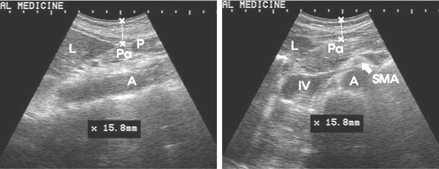
Ultrasonography findings on geonri-hyeol(CV11,
建里穴) of 42kg–150cm woman
(a) Longitudinal US image shows 15.8mm depth from epidermis to surface of pancreas body, (b) Transverse US image shows 15.8mm depth from epidermis to surface of pancreas body.
L : left lobe of liver, Pa: pancreas, IV: inferior vena cava. A: abdominal aorta, SMA: superior mesenteric artery
Fig. 27.
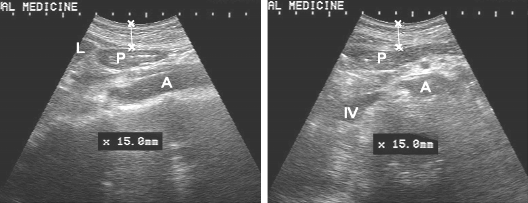
Ultrasonography findings on the hawan-hyeol(CV10,
下脘穴) of 42kg–150cm woman
(a) Longitudinal US image shows 15.0mm depth from epidermis to surface of pylorus.
(b) Transverse US image shows 15.0mm depth from epidermis to surface of pylorus.
L : left lobe of liver, P: pylorus, IV: inferior vena cava. A: abdominal aorta
참고문헌
1.. 衰秉哲譯. 今釋黃帝內經 靈福. 서울: 成輔社;1995. p. 168–9. p. 227p. 290–2. p. 293–5. p. 360–372. p. 623
2.. Heo Y. Technology Trends on Digital X-ray Diagnostic Imaging Devices. Journal of the Electrical World. 2008; 381:18–24.
3.. 司馬 遷. 史記. 合北: 安業書局有限公司;1990. p. 2788
4.. 정제. 최신침구학. 서울: 成輔社;1979. p. 24
5.. 衰秉哲譯. 今釋黃帝內經 素問. 서울: 成輔社;1994. p. 171–2. p. 350p. 430
6.. 김 규열, 배 병철. 한의학개론. 서울: 成輔社;2010. p. 19
7.. Park TH, Lee CW. Studies on Recognition of Human Anatomy and Development of Anatomy knowledge in the Orient. The Journal of Daejeon Korean Medicine. 1992; 1:1. 297–318.
8.. 김 용진, 윤 창렬. 난경 연구집성. 서울: 주민출판사;2002. p. 571p. 649p. 654–5.
9.. 심 찬섭. 복부초음파진단학. 서울: 여문각;2002. 194:p. 386
10.. 集 元方. 諸病源候論. 北京: 人民衛生出版社;1982. p. 157p. 194p. 228
11.. Sung MG, Jeong CH. A Comparative Study on Sinhyeongjangbudo. The Journal Of Korean Medical Classics. 2008; 21:3. 165–75.
12.. 홍 원식. 중국의사학. 서울: 동양의학연구원;1987. p. 150–1.
13.. Sm KH. The Concept of Anatomy and Body Perspective in Ancient China. Institute for History of Medicine, Yonsei University. 2012; 15:2. 29–61.
14.. 곽 동렬, 김 재원. 醫林改錯評譯. 서울: 成輔社;1998. p. 27p. 35p. 129
15.. Kim SM. History of Human Anatomy in Korean Medicine. The Journal of Daejeon Korean Medicine. 1995; 4:1. 461–65.
16.. 송 한덕. 초음파진단의 이해. 서울: 군자출판사;1995. p. 2–4. p. 150
17.. Kim BS, Kang JS. Study on Dermatology in Oriental Medicine. Korean Journal of Oriental Physiology and Pathology. 2002; 16:6. 1110–16.
19.. 이 활. 경동맥초음파. 서울: 대한의학서적;2011. p. 22p. 46p. 72
20.. Bianchi S, Martinoli C. Ultrasound of the Musculoskeletal System. New York: Springer;2007. p. 22–3.
21.. Cho MR, Kim MS, Ryu CR, Choi CH, Jang KS, So CH, et al. The quantitative study on the Renying.Qi mouth comparison pulse diagnosis. The Journal of Korean Acupuncture & Moxibustion Society. 2002; 19:2. 149–63.
22.. 전국한의과대학 피부외과학 교재편찬위원회. 한방피부외과학. 부산광역시: 도서출판 선우;2007. p. 35p. 192p. 201p. 211–22. p. 325–39.
23.. 신 소화관초음파 아틀라스. 문영수. 서울: 한국 의학;2009. p. 60p. 66–7. p. 93
24.. Hong SP. Functional Dyspepsia and Gastric Motility Disorders. Journal of Neurogastroenterology and Motility. 2000; 6:2. 241–7.
25.. 전국한의과대학 간계내과학교수. 간계내과학. 서울: 동양의학연구원;1989. p. 88
26.. 전국한의과대학 비계내과학교수. 비계내과학. 서울: 군자출판사;2008. p. 150p. 169p. 189–96.
27.. 두 호경. 동의신계내과학. 서울: 동양의학연구원;1986. p. 61p. 186
28.. Moon JJ, Vartan FR. Comparative Study on the Human and Equine Meridians. The Journal of Kyunghee Korean Medicine. 1980; 3:0. 123–74.
29.. 小川 鼎三. 医学用語の起り. 東京: 東京書籍株式會社;1990. p. 208–11.