


Introduction
Among cognitive impairment symptoms, dementia is commonly observed in the elderly, particularly those over the age of 80 years. Alzheimer’s disease (AD) is one of the most important causes of dementia, and cerebrovascular disease (CVD) also generally occurs in the elderly1). In addition, the incidence of vascular disease is much higher in Alzheimer’s patients who are clinically diagnosed with dementia, indicating that various vascular risk factors are involved in the pathogenesis of dementia1) Therefore, the management for cardiovascular and cerebrovascular risk factors in the early stage of dementia has been importantly considered to delay and suppress the dementia.2) In the early stages of dementia, both neuroinflammation and generation of reactive oxygen species are involved in the pathological changes by oxidative-induced inflammatory damage to small blood vessels.3) In addition, ischemia activates amyloid processing enzymes and other proinflammatory factors, resulting in confusion of the neuronal function.4) Moreover, because vascular dementia shares risk factors with CVD, such as cerebral infraction, antithrombotic drugs, such as platelet-aggregation inhibitors, are prescribed in the clinical setting to prevent and treat vascular dementia.5) Furthermore, EGb 761, ginkgo biloba extract and Woo-Gui-Um extract are known to be effective for preventing dementia.6–8) However, the correlation between these drugs and dementia prevention remains to be clarified. The application of ferric chloride (FeCl3) is commonly used to cause vascular injury and thrombosis in an animal model, which model has been known to be sensitive to anticoagulant and antiplatelet drugs.9) FeCl3 causes oxidative stress, which leads to lipid peroxidation and destruction of endothelial cells, through inducing the generation of free radicals and occlusive thrombus formation.4,10) Based on this fact, FeCl3-induced arterial injury model has been implemented as the primary method to develop new drug candidates for dementia prevention.11) Previous reports have verified the efficacy of merely antithrombotic drugs, such as clopidogrel,12) but there are no studies screening the efficacy of herbal prescriptions. For example, ginkgo biloba, green tea, and flavonoids showed a therapeutic effect in patients with degenerative and vascular diseases.13, 14) Therefore, in this study, we primarily examined the efficacy of herbal prescriptions for blood vessel injury, selected prescriptions through qualitative assessment of research papers for herbal medicines specifically prescribed for treatment of dementia,15) and then established the basis of efficacy of the new potential candidates. Taken together, we evaluated the potential effect of 8 types of herbal prescriptions in FeCl3-induced arterial thrombosis animal model in vivo, and then found that CMT and LMK02 may be the most promising candidates for the prevention and treatment of vascular dementia.
Materials and methods
1. Materials and reagents
Yokukansan, Chotosan, Oren-gedoku-to and Toki-shakuyaku-san were purchased from Tsumura & Co (Tokyo, Japan). modified Jangwonhan 01 (LMK01), modified Jangwonhan 02 (LMK02) and modified Jangwonhan 03 (LMK03) were supplied by Hanpoong Pharm & Food Co., Ltd (Jeonju, Korea). Table 1 shows the composition of the herbal prescriptions. Isofluran/foran solution (Choongwae Pharmaceutical Co., Seoul, Korea) was prepared by mixing oxygen (Shinheung Oxygen, Daejeon, Korea) and nitrogen (Shinheung Oxygen). Ferric chloride and other chemicals were purchased from Sigma-Adrich (St Louis, MO, USA). A small animal anesthesia machine (VMC Anesthesia Machine, Midmark International, Versailles, OH, USA) was used. Laser Doppler flowmetry (LDF; BFL21, Transonic Instrument, NY, USA) including a miniature probe was used (Powerlab/8sp, ADInstruments Pty Ltd, Castle Hill, NSW, Australia).
2. Preparation of Chong-myung-tang (CMT) extract
To prepare CMT extract, 3 types of herbs were purchased from Kwangmyungdang Medicinal Herbs in Ulsan, Korea, in 2011. All herbs were identified by Professor Bang Yeon Hang from the College of Pharmacy, Chungbuk National University and deposited at the herbarium of the Herbal Medicine Research Division, Korea Institute of Oriental Medicine (KIOM, Korea). The ingredients of CMT were Acorus gramineus Soland (Acori Rhizoma), Polygala tenuifolia Willdenow (Polygalae Radix) and Poria cocos Wolf (Polia). They were mixed in a ratio of 1:1:1 to reach a total weight of 4.5 kg. The mixture was extracted twice by refluxing in distilled water (1:10 w/v) for 2 hours below 100°C. After filtration, all extracts were concentrated with a rotary evaporator and were then freeze-dried to yield an extract powder (390 g, yield 8.6% w/w).
3. Experimental animals
All experimental procedures were approved by the Animal Care and Use Committee of the Korea Institute of Oriental Medicine (Approval no. 11-090). Male Sprague-Dawley (SD) rats, 8–10 weeks old, weighting 230–250 g (Orient Bio Inc., Seongnam, Korea) were used for the FeCl3-induced carotid arterial injury model. Animals were housed in polyethylene cages in an animal room with a controlled temperature at 24 ± 2°C, a constant humidity of 50 ± 10%, and a 12-h light-dark cycle. Animals had free access to a standard diet and tap water ad libitum.
4. FeCl3-induced carotid arterial thrombosis
The FeCl3-induced carotid arterial injury model was used as previously described with minor modifications.10,16,17) Briefly, male SD rats were anaesthetized with 2% isoflurane in a mixture of 70% nitrous oxide and 30% oxygen, using facemask anaesthesia, which was continued throughout the surgical procedure. Body temperature was maintained at 37 ± 2°C with a heating pad throughout surgery (LCI, Seoul, Korea). An incision of the skin was made directly on top of the right common carotid artery. Carotid blood flow was measured with a miniature Doppler flow probe (BFL21, Transonic Instrument, Ithaca, NY). Thirty minutes prior to 35% FeCl3 treatment, rats were injected with either saline or herbal prescriptions (100 mg/kg, i.p.). A small piece of filter paper, soaked in FeCl3 solution (35%, w/v, Sigma-Aldrich), was then applied topically to the carotid artery of these SD rats for 3 min and then removed. To measure the occlusion time in the carotid artery, carotid blood flow was continuously monitored for 30 min after FeCl3 application. At the end of the experiment, the arterial including thrombus was excised and weighed.
5. Histological analysis
After the FeCl3-induced carotid arterial injury, carotid artery samples were fixed in 10% buffered formalin and then embedded in paraffin for sectioning. Several sections obtained from each artery were excised and fixed in 10% PBS-buffered formal in for 24 hours. Following paraffin embedding and sectioning (4 μm), the tissues were stained with hematoxylin and eosin (H&E), Masson’s trichrome stain. The collagen fibers will be stained blue, the nuclei will be stained black and the background will be stained red.18) The stained sections were examined at a magnification of 40x to study thrombus formation and vascular wall structure. Photomicrographs were taken using an Olympus BX51 photomicroscope (Olympus, Tokyo, Japan).19) Quantitative analysis of distribution of collagen fiber staining was measured using Meta Imaging Series 7.7. (Molecular Devices, NY, USA).
6. Statistical analysis
All statistical analyses were performed using SPSS version 12.0 (SPSS Inc., Chicago, IL, USA) and R package version 2. 12.0, with data shown as mean ± standard deviation (SD). Statistical evaluations of the data were performed by one-way ANOVA followed by Duncan’s post-hoc test. Values of P<0.05 were considered as statistically significant.
Results
1. In vivo antithrombotic effects of herbal prescriptions in the FeCl3-induced arterial thrombosis model
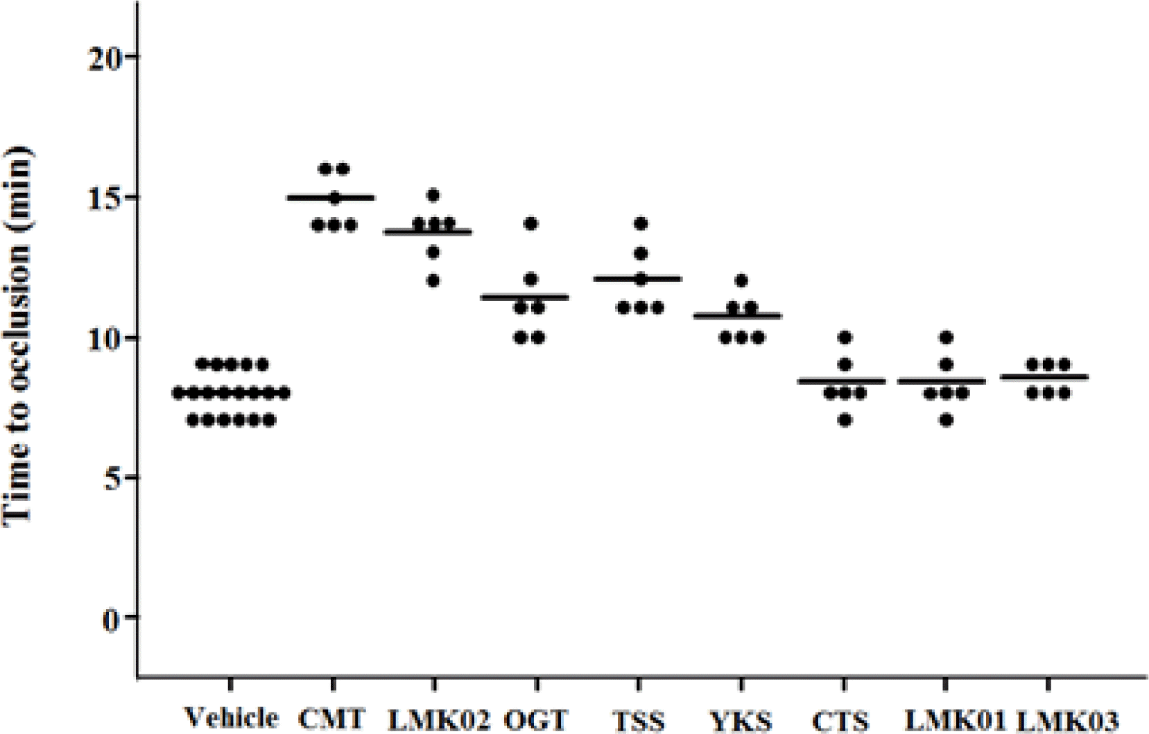
To evaluate antithrombotic effects of 8 types of herbal prescriptions in vivo, the FeCl3-induced arterial thrombosis model was established11) and used for this study. We observed formation of an occlusive, platelet-rich thrombus within 30 min after FeCl3 injury. Figure 1 shows the effect of the herbal prescriptions on time to occlusion (TTO) in arterial thrombosis model. The mean TTOs for Chong-myung-tang (CMT), LMK02, Toki-shakuyaku-san (TSS), Oren-gedoku-to (OGT), and Yokukansan (YKS) (14.83 ± 0.98, 13.67 ± 1.03, 12.00 ± 1.26, 11.33 ± 1.51 and 10.67 ± 0.82 min, respectively) were significantly longer than that in the vehicle group (8.06 ± 0.77 min; P<0.001, Table 2). In particular, CMT remarkably delayed the TTO compared with LMK02 (P<0.05). However, Chotosan (CTS), LMK01, and LMK03 did not delay the TTO. In addition, treatment with CMT, LMK02, and OGT meaningfully reduced thrombus weight (0.63 ± 0.01, 0.66 ± 0.02, 0.67 ± 0.01 mg/mm, respectively) compared with vehicle (0.78 ± 0.03 mg/mm; P<0.001, Table 2). Taken together, these results suggest that CMT, LMK02, and OGT among the investigated herbal prescriptions may be the outstanding candidates to suppress FeCl3-induced thrombosis.
2. Histological analysis for vascular injury
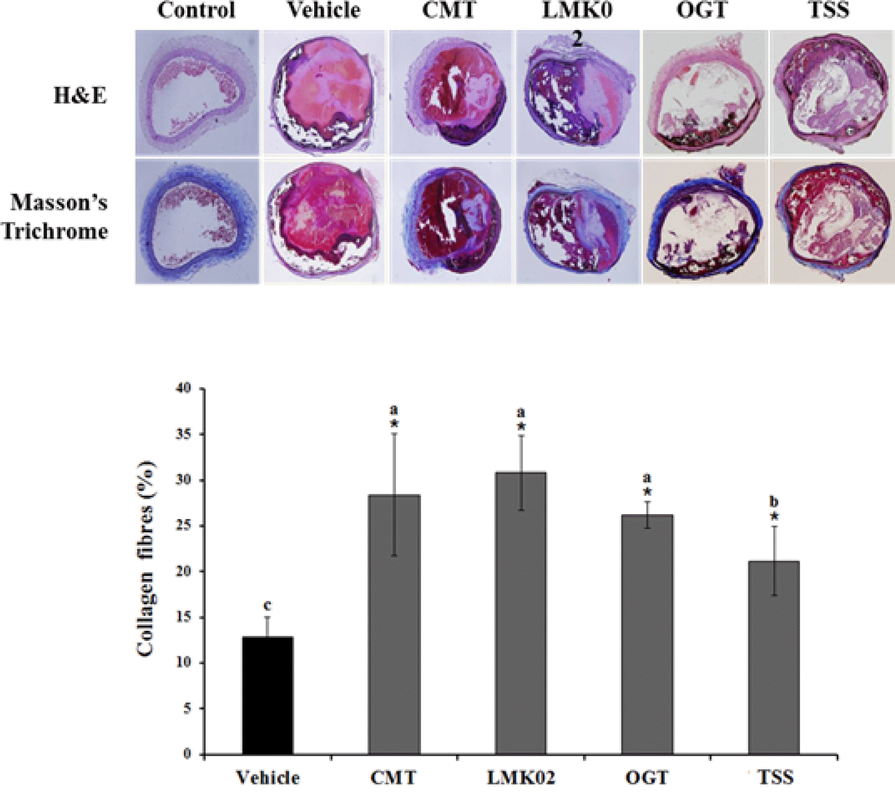
High concentrations of FeCl3 induce vascular injury, such as endothelial denudation, collagen exposure, and thrombus formation.20) Hematoxylin and eosin (H&E) staining was performed to investigate histological differences between normal and thrombotic vessels. Masson’s trichrome staining was implemented to quantify areas containing fibrillar collagen. Fig. 2 shows representative photomicrographs of H&E or Masson’s trichrome staining in the carotid artery after injury with FeCl3. The collagen fibers damage was detected in FeCl3-treated rats (12.80 ± 2.22%, Fig. 2), but were significantly ameliorated in the CMT-treated group (28.40 ± 2.22%, P<0.001), LMK02-treated group (30.79 ± 4.07%, P<0.001), OGT-treated group (26.20 ± 1.48%, P<0.05) and TSS-treated group (21.14 ± 3.78%, P<0.05) compared with the vehicle-treated group. In the previous study, collagen fibers damage was not observed in the carotid artery of saline-treated rats. Furthermore, in Masson’s trichrome staining, collagen fibers in the vessels showed histological changes that typically occur in the carotid arteries of SD rats after FeCl3 injury. The distribution of collagen fibers was decreased in the vehicle-treated carotid arteries by FeCl3 treatment (Fig. 2). However, CMT and LMK02 restored the decreased distribution of collagen fibers by more than 50% protection of FeCl3-induced damage of collagen fibers, suggesting that CMT and LMK02 may be excellent activity for restoring the reduced distribution of collagen fibers induced by FeCl3 treatment.
Discussion
Alzheimer’s disease has received attention with the aging of global populations for several decades. There is a hypothesis that amyloid-♌ (A ♌) is the primary cause of dementia; drug development is carried out on the basis of this hypothesis.21) Recent advances have drawn attention to strategies of multi-target drug development instead of one-target drug development. Conventional herbal medicines have multi-target specificity. For these reasons, it is important to examine the potential efficacy of herbal medicines and the concept of new drug treatment utilizing traditional herbal medicines needs to be applied.22) We used an animal model as an in vivo assay system to screen the efficacy of herbal prescriptions that were applied to prevent vascular injury, such as vascular oxidation and inflammation. FeCl3 induces endothelial denudation through oxidative stress, exposing the collagen matrix for the site of adhesion and activation of circulating platelets.23) The FeCl3-induced arterial thrombosis model affects platelet aggregation and activation, collagen exposure, and vascular injury.9) Although this study did not examine how any single compound in herbal prescriptions consisting of many types of compounds was effective in FeCl3-induced carotid arterial injury, we found that CMT and LMK02 extract significantly ameliorated TTO and thrombus weight and altered distribution of collagen fibers in the FeCl3-induced arterial thrombosis model. CMT is a traditional herbal prescription in Korea, and is widely used as a remedy for amnesia to ameliorate learning and memory. According to Lee et al. (2010), 200 mg/kg/day of CMT significantly restored memory impairment induced by scopolamine in mice.24) In addition, Kim et al. (1999) demonstrated that CMT has an inhibitory activity of tumor necrosis factor-α production in mouse astrocytes stimulated with lipopolysaccharide plus substance P.25) However, a protective effect for CMT on vascular injury has not been previously reported. We found, for the first time, that CMT had the greatest efficacy among our herbal prescriptions used. In our other study, the effects of the ingredients which compose CMT were investigated in vascular injury, and we found that Acorus gramineus among the ingredients showed markedly preventive activity (data not shown); therefore, this needed to be verified by additional research. In addition, LMK02 and LMK03 modified from Jangwonhwan (LMK01) have been reported to reduce Aβ levels in the Tg-APPswe/PS1dE9 mouse model of Alzheimer’s disease.26,27) In this study, LMK02 showed a preventive effect, similar to CMT in the FeCl3-induced arterial thrombosis model. However, LMK03 and LMK01 were not effective. It is presumed that red ginseng and Acorus gramineus, ingredients which appear in LMK02, have antithrombotic effect, but efficacy evaluation for all ingredients is necessary.28) Although the correlation between thrombus and dementia prevention has not been precisely clarified, alternative medications in vascular injury are anticipated to contribute to relieving symptoms in dementia patients and restoring vascular cognitive impairment. Furthermore, it is necessary to investigate the mechanism of action of CMT and LMK02 in the investigation of new drugs for the prevention of vascular dementia or arterial thrombosis. CMT and LMK02 were first verified as effective in vivo antithrombotic drugs in our study. In conclusion, our results demonstrated protective effects of both CMT and LMK02 on an animal thrombosis model. This indicates that CMT and LMK02 are potential therapeutic agents for thrombosis and vascular injury. Further study is required to determine the mechanisms of action of ingredients to aid in the discovery of new drugs for the prevention of vascular dementia or arterial thrombosis.