Anti-inflammatory Effect of Cornus Officinalis fruit extract and Cornus Officinalis Fruit Cheonghyeol Plus in Human Umbilical Vein Endothelial Cell

Article information

J Korean Med. 2022;43(3):106-121
Publication date (electronic) : 2022 September 1
doi : https://doi.org/10.13048/jkm.22034
Dept. of Internal Medicine, College of Korean Medicine, Daejeon University
Correspondence to: Yoonsik Kim, Oriental Medicine Department of Daejeon University Cheonan Oriental Medicine Hospital, Daejeon University, Notaesan-ro 4-gil, Seobuk-gu, Cheonan-si, Chungcheongnam-do, Tel: +82-41-521-7536, Fax: +82-41-521-7007, E-mail: yoonsik@dju.kr
Received 2022 July 20; Revised 2022 August 10; Accepted 2022 August 16.

Abstract

Objectives

The purpose of this study was to investigate the anti-inflammatory effect of Cornus Officinalis fruit extract(CE) and Cornus Officinalis Fruit Cheonghyeol Plus(CCP) in Human Umbilical Vein Endothelial Cell.

Methods

We measured cell viability of CE, CCP and treated HUVEC with TNF-α. We measured the mRNA expression levels of KLF2, eNOS, MCP-1, ICAM-1, VCAM-1, the protein expression levels of KLF2, eNOS, MCP-1, ICAM-1, VCAM-1, and the protein phosphorylation level of ERK, JNK, p38 and the biomarker expression levels of MCP-1, ICAM-1, VCAM-1.

Results

1.CE incresed the mRNA, protein expression levels of KLF2, eNOS at concentrations of 100μg/ml compared to the control group. CE decresed the mRNA, protein and biomarker expression levels of MCP-1,ICAM-1,VCAM-1 at concentrations of 100μg/ml compared to the control group. CE decresed the protein phosphorylation level of p38 at concentrations of 100μg/ml compared to the control group. 2. CCP incresed the mRNA, protein expression levels of KLF2, eNOS at concentrations of 100μg/ml or more compared to the control group. CCP decresed the mRNA, protein and biomarker expression levels of MCP-1, ICAM-1, VCAM-1 at concentrations of 100μg/ml or more compared to the control group. CCP decresed the protein phosphorylation level of ERK at concentrations of 100μg/ml or more, p38 at concentrations of 200μg/ml or more, and JNK at concentrations of 400μg/ml compared to the control group.

Conclusions

These results present that CE and CCP has anti-inflammatory effect in HUVEC. So, it could help treat or prevent inflammation in vein caused by dyslipidemia and contribute prevention of cardiovascular and cerebrovascular cerebrovascular diseases.
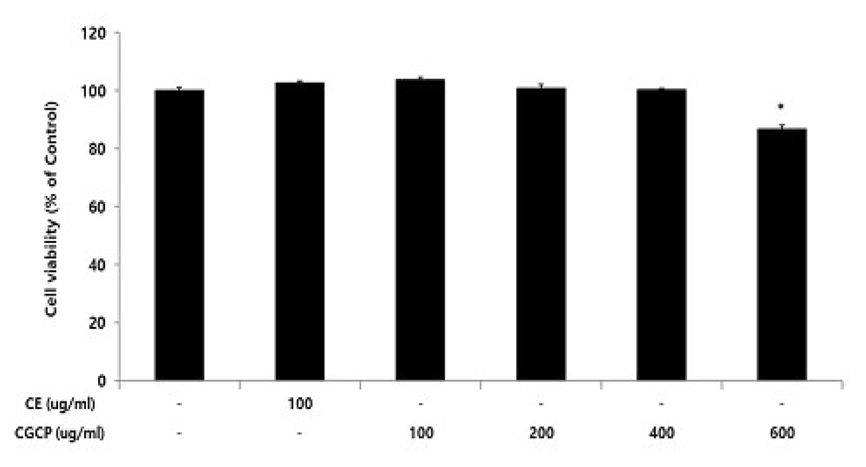
Fig. 1

Cell viability of CE or CCP in HUVEC

Cell viability were calculated as percentage relative to the control and the result were given by the mean±standard error of mean from three independent experiments(Significance of results, *; p<0.05 compared to control).

Fig. 2

Effect of CE or CCP on KLF2(A), eNOS(B), MCP-1(C), ICAM-1(D), VCAM-1(E) mRNA expression level in HUVEC

HUVEC were treated CE(100 ug/ml or CCP(100, 200, and 400 ug/ml), with TNF-α 10 ng/ml for 12hour. The mRNA expression level were measured using a real-time PCR(qPCR). The result were given by the mean±standard error of mean from three independent experiments(Significance of results, *; p<0.05, **; p<0.01, ***; p<0.001 compared to control, +++; p<0.001 compared to normal).
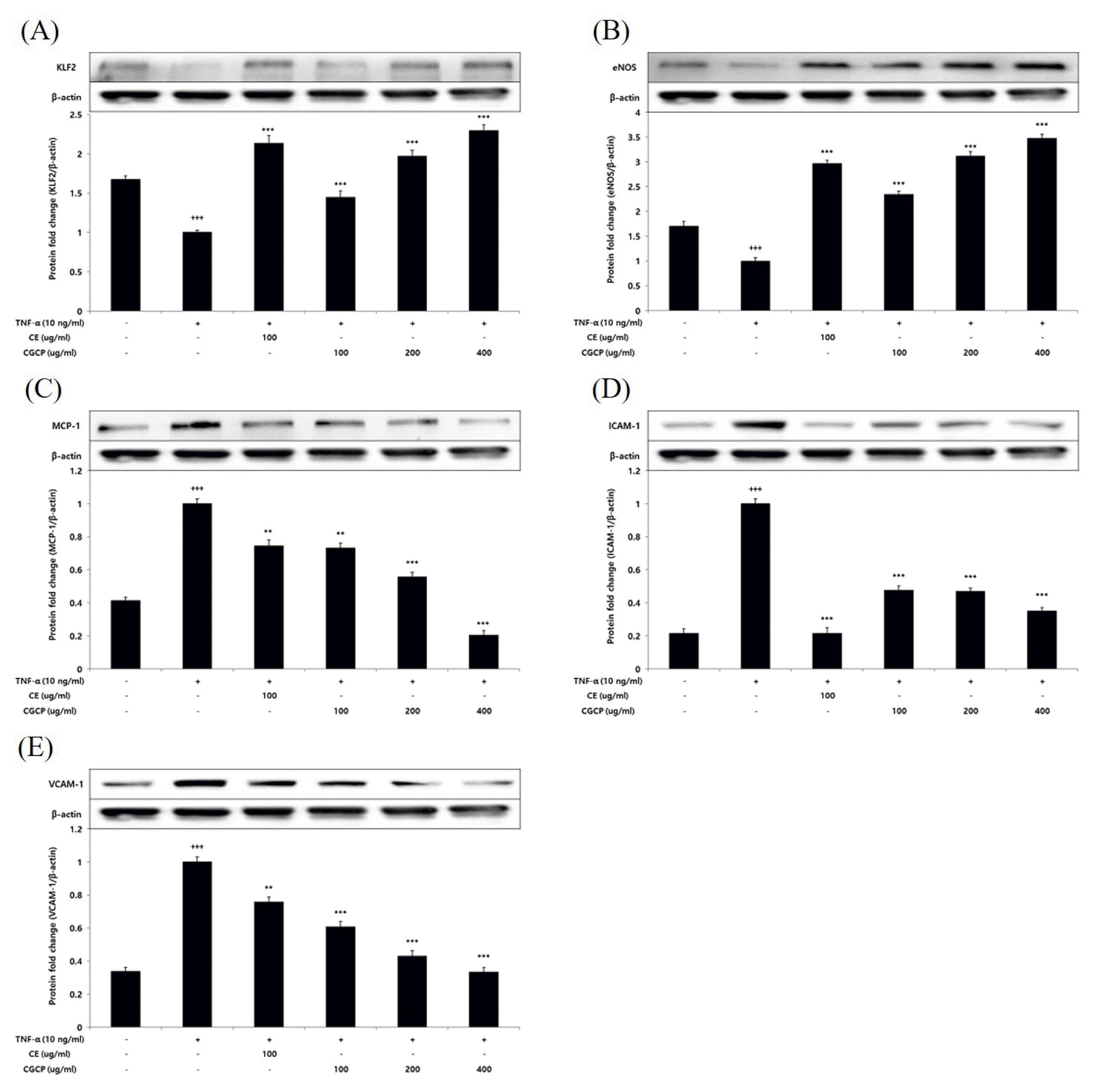
Fig. 3

Effect of CE or CCP on KLF2(A), eNOS(B), MCP-1(C), ICAM-1(D), VCAM-1(E) protein expression level in HUVEC

HUVEC were treated CE(100ug/ml or CCP(100, 200, and 400ug/ml), with TNF-α 10 ng/12 hour. The protein expression level were measured by a western blot. The result were presented by the mean±standard error of mean from three independent experiments(Significance of results, *; p<0.05, **; p<0.01, ***; p<0.001 compared to control, +++; p<0.001 compared to normal).
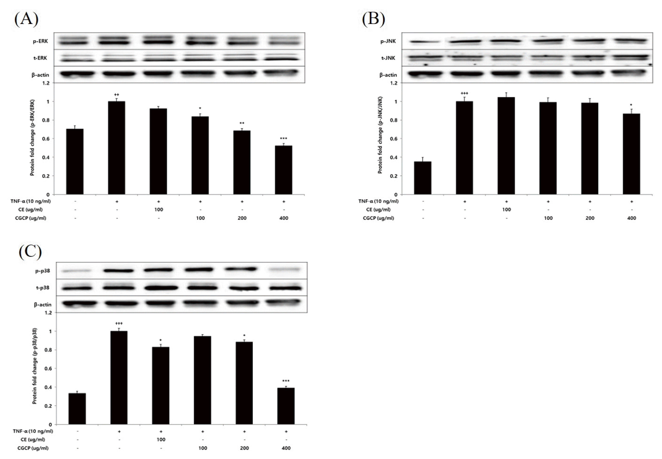
Fig. 4

Effect of CE or CCP on ERK(A), JNK(B), p38(C) protein phosphorylation level in HUVEC

HUVEC were treated CE(100ug/ml or CCP(100, 200, and 400ug/ml), with TNF-α 10 ng/12 hour. The protein phosphorylation level were measured by a western blot. The result were presented by the mean±standard error of mean from three independent experiments(Significance of results, *; p<0.05, **; p<0.01, ***; p<0.001 compared to control, +++; p<0.001 compared to normal).
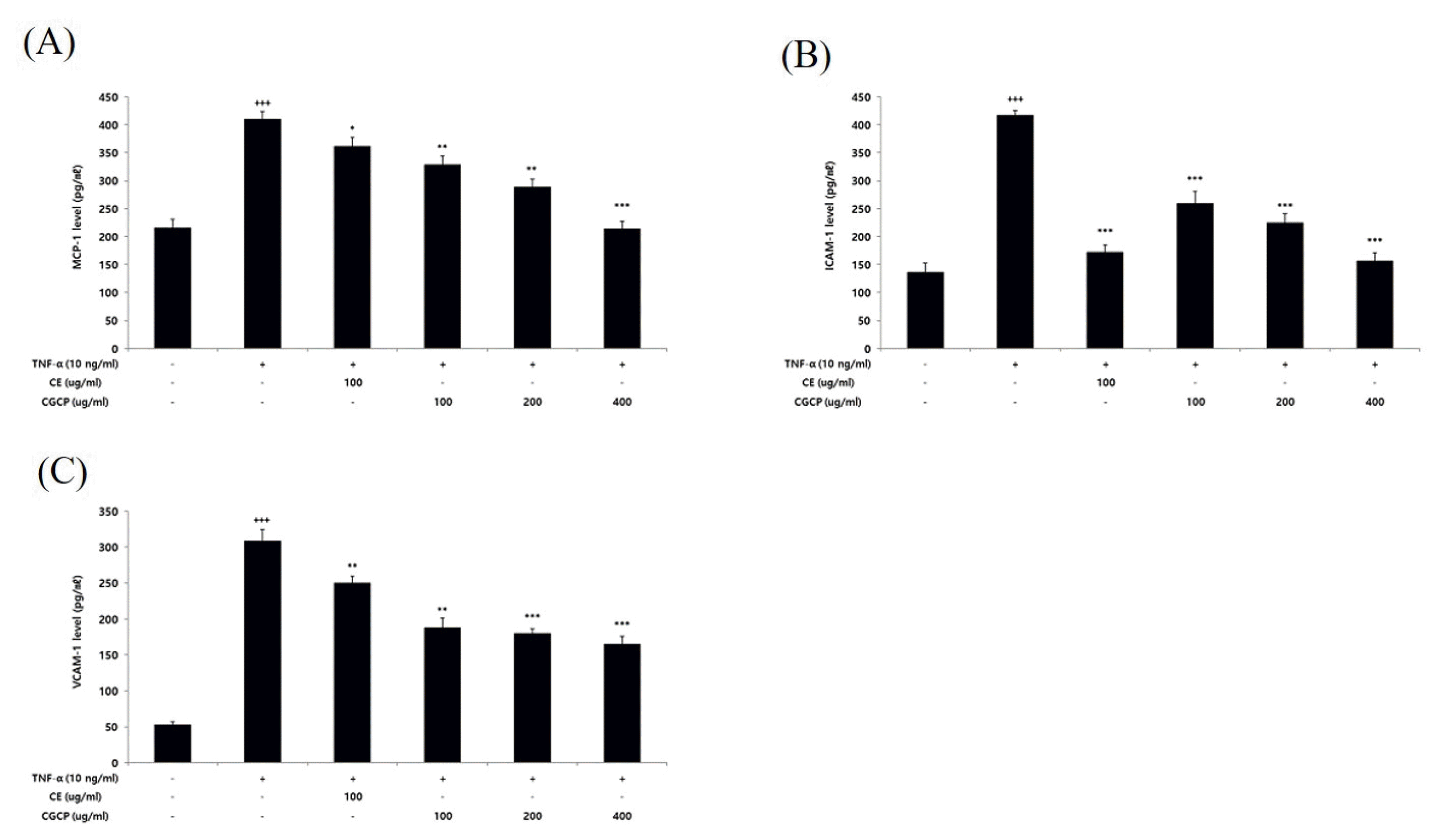
Fig. 5

Effect of CE or CCP on MCP-1(A), ICAM-1(B), VCAM-1(C) level in HUVEC

HUVEC were treated CE(100ug/ml or CCP(100, 200, and 400ug/ml), with TNF-α 10 ng/12hour. MCP-1 level was measured by ELISA kit. The result were presented by the mean±standard error of mean from three independent experiments(Significance of results, *; p<0.05, **; p<0.01, ***; p<0.001 compared to control, +++; p<0.001 compared to normal).

CE : Cornus Officinalis fruit extract

CCP : Cornus Officinalis Fruit Cheonghyeol Plus

HUVEC : Human Umbilical Vein Endothelial Cell

KLF : Kruppel-like factor 2

eNOS : Endothelial nitric oxide synthase

MCP-1 : Monocyte chemoattractant protein-1

ICAM : Intercellular Adhesion Molecule -1

VCAM : Vascular Cell Adhesion Molecule -1

TNF : Tumor necrosis factor-α

ERK : Extracellular signal-regulated kinase

JNK : Jun N-terminal kinase

Real-Time PCR Primer Sequences

References

1. Oh B.H.. 2003;New Concepts in the pathogenesis and Progression of Atherosclerosis. Medical postgraduates 31(4):179–183.
2. Statistics Korea. 2021. 2020 Death statistics : Korea Korea development institute; Available from: URL: http://kostat.go.kr/portal/korea/kor_nw/1/6/2/index.board?bmode=read&bSeq=&aSeq=403046&pageNo=1&rowNum=10&navCount=10&currPg=&searchInfo=&sTarget=title&sTxt= .
3. Jeong H.S.. 2019. Risks and Management of Dyslipidemia. Public health weekly report 2019. 12(37)1416–1211. Available from: URL: https://www.kdca.go.kr/board/board.es?mid=a20602010000&bid=0034&list_no=364839&act=view .
4. Bae J.H., Park J.S., Hond G.R., Shin D.G., Kim Y.J., Shim B.S.. 2008;Correlation between inflammatory markers and the progression of atherosclerosis in patients with coronary artery disease. The Korean Journal of Medicine 74(1):51–58.
5. Stenvinkel P.. 2003;Interactions betwen inflammation, oxi dative stres, and endothelial dysfunction in end-stage re nal disease. J Ren Nutr 13(2):144–148. https://doi.org/10.1053/jren.2003.50018 .
6. Wolf D., Ley K.. 2019;Immunity and Inflammation in Atherosclerosis. Circ Res 124(2):315–327. https://doi.org/10.1161/CIRCRESAHA.118.313591 .
7. Glass C.K., Witztum J.L.. 2001;Atherosclerosis.The road ahead. Cell 104(4):503–516. https://doi.org/10.1016/s0092-8674(01)00238-0 .
8. Seo D.H., Joo I.H., Kim D.H.. 2018;Effect of ChungHuyl-Plus on inflammatory factors in Human Umbilical Vein Endothelial Cells (HUVECs). J Haehwa Medicine 27(2):11–20.
9. Oh J.M., Cho H.K., Yoo H.R., Kim Y.S., Seol I.C.. 2016;A Case Report of the Beneficial Effects of Chunghyul-Plus in Dyslipidemia Patients. The Journal of the Society of Stroke on Korean Medicine 17(1):55–66.
10. Park E.B., Kim H.S., Shin S.Y., Ji I.A., Kim J.H., Kim S.G., et al. 2021;Antioxidative Activity of Cornus officianalis Extracts Obtained by Four Different Extraction Techniques. Journal of Life Science 22(11):1507–1514. http://dx.doi.org/10.5352/JLS.2012.22.11.1507 .
11. Kim Y.J., Son D.Y.. 2016;Antioxidant activity and suppression of pro-inflammatory mediator of Corni fructus extracts in activated. Korean J Food Preserv 23(6):876–882. https://doi.org/10.11002/kjfp.2016.23.6.876 .
12. Shin J.H., Cha G.Y., Kim H.J., Hwang J.H., Han K.H., Seo H.J., et al. 2009;Exmination of Anti-Obesity Effect of RegionalSpecial Natural Products of Anthrisci radix, Psoraleaesemen, Siegesbeckiae herba and Corni fructus. KSBB Journal 24(6):549–555.
13. Yamabe N., Noh J.S., Park C.H., Kang K.S., Shibahara N., Tanaka T., et al. 2010;Evaluation of loganin, iridoid glycoside from Corni Fructus, on hepatic and renal glucolipotoxicity and inflammation in type 2 diabetic db/db mice. European Journal of Pharmacology 648(13):179–187. https://doi.org/10.1016/j.ejphar.2010.08.044. Epub 2010 Sep 15.
14. Jiang Z.Q., Li Y., Jiang L.H., Gu H., Wang M.Y.. 2013;Hepatoprotective effects of extracts from processed corni fructus against D-galactose-induced liver injury in mice. Zhong Yao Cai 36(1):85–89.
15. Ross R.. 1993;The pathogenesis of atherosclerosis: A perspective for the 1990s. Nature. 362(6423):801–809. https://doi.org/10.1038/362801a0 .
16. Committee of Clinical Practice Guideline of the Korean Society of Lipid and Atherosclerosis (KSoLA). 2018. Korean Guidelines for the Management of Dyslipidemia 4th edth ed. Seoul: p. 31–44.
17. Li J.J., Fang C.H.. 2004;Atheroscleritis is a more rational term for the pathological entity currently known as atherosclerosis. Med Hypotheses 63(1):100–102. https://doi.org/10.1016/j.mehy.2004.01.029 .
18. Bergheanu S.C., Bodde M.C., Jukema J.W.. 2017;Pathophysiology and treatment of atherosclerosis. Neth Heart J 25(4):231–242. https://doi.org/10.1007/s12471-017-0959-2 .
19. Yang Y.K.. 1991. Hwangjenegyung-yuckhe Seoul: Iljoong-sa.
20. Yang J.H., Yoo H.R., Kim Y.S., Seol I.C.. 2021;The Effect of Lonicera Japonica Thunberg on Inflammatory Factor Expression Associated with Atherosclerosis. Korean J. Orient. Int. Med 42(1):25–39. https://doi.org/10.22246/jikm.2021.42.1.25 .
21. Do H.J., Kim K.H., Oh T.W.. 2020;Anti-hyperlipidemic Effects of Scutellariae Radix, Aucklandiae Radix and Bupleuri Radix (SAB) extract in FL83B cells. Kor. J. Herbol 35(5):23–31. https://doi.org/10.6116/kjh.2020.35.5.23 .
22. Han B.H., Yoon J.J., Kim H.Y., Ahn Y.M., Hong M.H., Son C.O., et al. 2018;Inhibitory Effects of Ojeoksan on TNF-α-induced Vascular Inflammation in Human Umbilical Vein Endothelial Cells. Kor. J. Herbol 33(4):59–67. https://doi.org/10.6116/kjh.2018.33.4.59 .
23. Lee K.W., Cho H.K., Yoo H.R., Seol I.C.. 2018;The Effects of an Extract of Fermented Artemisiae Iwayomogii Herba, Curcumae Longae, Crataegi Fructus and Salviae Miltiorrhizae Radix on Anti-inflammation Associated with Dyslipidemia and Anti-oxidation in RAW264.7 and HUVEC Cells. J. Int. Korean Med 39(4):480–494. https://doi.org/10.22246/jikm.2018.39.4.480 .
24. Choi K.E., Seol I.C., Kim Y.S., Jo H.K., Yoo H.R.. 2016;Hypolipidemic and Anti-oxidant Effects of Chunghyl Plus in Type II Diabetic Mice Model. J Physiol & Pathol Korean Med 30(3):164–176. https://doi.org/10.15188/kjopp.2016.06.30.3.164 .
25. Lee J.H., Jang H.J., Kim K.S., Yang H.J., Lee J.Y., Na Y.C., et al. 2014;Effects of β-sitosterol derived from Artemisia capillaris on the activated human hepatic stellate cells and dimethylnitrosamine-induced mouse liver fibrosis. BMC Complement Altern Med 14(363):1–10. https://doi.org/10.1186/1472-6882-14-363 .
26. Oh D.Y., Kang D.S., Lee Y.G., Kim H.S.. 2019;Effects of Turmeric (Curcuma longa L.) on Blood Glucose and Lipid Metabolism Functional Improvement in STZ-induced Diabetic rats. J Environmental Science International 28(5):485–494. https://doi.org/10.5322/JESI.2019.28.5.485 .
27. Oh D.Y., Kim H.S.. 2019;Evaluation of Oxidation Inhibition and Nitrogen Oxide Scavenging Activity from Curcuma longa L. Extracts. J Kor. applied science and technology 36(1):13–22. https://doi.org/10.12925/jkocs.2019.36.1.13 .
28. Kwon S.H., Kim J.B.. 2010;Effects of Crataegii Fructus on the Diet-induced Hyperlipidemia in Rats. Korean J. Oriental Physiology & Pathology 24(1):67–73.
29. Lee S.E., Cho S.I.. 2015;Anti-inflammatory effects of Salviae Miltiorrhizae Radix extract on RAW264.7 cell. via anti-oxidative activities. Kor. J. Herbol 30(4):89–94. http://dx.doi.org/10.6116/kjh.2015.30.4.89 .
30. Zhang J., Liang R., Wang L., Yang B.. 2019;Effects and mechanisms of Danshen-Shanzha herb-pair for atherosclerosis treatment using network pharmacology and experimental pharmacology. J Ethnopharmacol, 2019 229(1):104–114. https://doi.org/10.1016/j.jep.2018.10.004 .
31. Packard R.R.S., Libby P.. 2008;Inflammation in atherosclerosis: from vascular biology to biomarker discovery and risk prediction. Clin Chem 54(1):24–38. https://doi.org/10.1373/clinchem.2007.097360 .
32. Onat D., Brillon D., Colombo P.C., Colombo P.C., Schmidt A.M.. 2011;Human Vascular Endothelial Cells: A Model System for Studying Vascular Inflammation in Diabetes and Atherosclerosis. Curr Diab Rep 11(3):193–202. https://doi.org/10.1007/s11892-011-0182-2 .
33. Bu D., Tarrio M., Grabie N., Zhang Y., Yamazaki H., Stavrakis G., et al. 2010;Statin-induced Kruppel-like factor 2 expression in human and mouse T cells reduces inflammatory and pathogenic responses. J Clin Invest 120(6):1961–1970. https://doi.org/10.1172/JCI41384. Epub 2010 May 3.
34. Boon R.A., Horrevoets A.J.. 2009;Key transcriptional regulators of the vasoprotective effects of shear stress. Hamostaseologie 29(1):39–43.
35. Yim C.Y.. 2010;Nitric oxide and cancer. Korean J Med 78(4):430–436.
36. Shin J.J., Lee S.Y., Lee S.H., Lee S.H., Suh J.K., Cho J.Y., et al. 1996;The Role and Localization of Nitric Oxide Synthase in Neurogenic Inflammation of the Rat Airways. Tuberculosis and Respiratory disease 43(3):420–432. https://doi.org/10.4046/trd.1996.43.3.420 .
37. Ramji D.P., Davies T.S.. 2015;Cytokines in atherosclerosis: Key players in all stages of disease and promising therapeutic targets. Cytokine Growth Factor Rev 26(6):673–685. https://doi.org/10.1016/j.cytogfr.2015.04.003. Epub 2015 May 12.
38. Kim E.K., Choi E.J.. 2015;Compromised MAPK signaling in human diseases: an update. Arch Toxicol 89(6):867–882. https://doi.org/10.1007/s00204-015-1472-2. Epub 2015 Feb 18.
39. Ryu I.H., Cho H.B., Kim S.B., Seo Y.J.. 2011;The inhibitory effect of Picrasmae Lignum on inflammatory responses. J Kor. Medicine Obstetrics and Gynecology 24(1):1–14. https://doi.org/10.15204/jkobgy.2011.24.1.001 .

Article information Continued

Fig. 1

Cell viability of CE or CCP in HUVEC

Cell viability were calculated as percentage relative to the control and the result were given by the mean±standard error of mean from three independent experiments(Significance of results, *; p<0.05 compared to control).

Fig. 2

Effect of CE or CCP on KLF2(A), eNOS(B), MCP-1(C), ICAM-1(D), VCAM-1(E) mRNA expression level in HUVEC

HUVEC were treated CE(100 ug/ml or CCP(100, 200, and 400 ug/ml), with TNF-α 10 ng/ml for 12hour. The mRNA expression level were measured using a real-time PCR(qPCR). The result were given by the mean±standard error of mean from three independent experiments(Significance of results, *; p<0.05, **; p<0.01, ***; p<0.001 compared to control, +++; p<0.001 compared to normal).

Fig. 3

Effect of CE or CCP on KLF2(A), eNOS(B), MCP-1(C), ICAM-1(D), VCAM-1(E) protein expression level in HUVEC

HUVEC were treated CE(100ug/ml or CCP(100, 200, and 400ug/ml), with TNF-α 10 ng/12 hour. The protein expression level were measured by a western blot. The result were presented by the mean±standard error of mean from three independent experiments(Significance of results, *; p<0.05, **; p<0.01, ***; p<0.001 compared to control, +++; p<0.001 compared to normal).

Fig. 4

Effect of CE or CCP on ERK(A), JNK(B), p38(C) protein phosphorylation level in HUVEC

HUVEC were treated CE(100ug/ml or CCP(100, 200, and 400ug/ml), with TNF-α 10 ng/12 hour. The protein phosphorylation level were measured by a western blot. The result were presented by the mean±standard error of mean from three independent experiments(Significance of results, *; p<0.05, **; p<0.01, ***; p<0.001 compared to control, +++; p<0.001 compared to normal).

Fig. 5

Effect of CE or CCP on MCP-1(A), ICAM-1(B), VCAM-1(C) level in HUVEC

HUVEC were treated CE(100ug/ml or CCP(100, 200, and 400ug/ml), with TNF-α 10 ng/12hour. MCP-1 level was measured by ELISA kit. The result were presented by the mean±standard error of mean from three independent experiments(Significance of results, *; p<0.05, **; p<0.01, ***; p<0.001 compared to control, +++; p<0.001 compared to normal).

CE : Cornus Officinalis fruit extract

CCP : Cornus Officinalis Fruit Cheonghyeol Plus

HUVEC : Human Umbilical Vein Endothelial Cell

KLF : Kruppel-like factor 2

eNOS : Endothelial nitric oxide synthase

MCP-1 : Monocyte chemoattractant protein-1

ICAM : Intercellular Adhesion Molecule -1

VCAM : Vascular Cell Adhesion Molecule -1

TNF : Tumor necrosis factor-α

ERK : Extracellular signal-regulated kinase

JNK : Jun N-terminal kinase
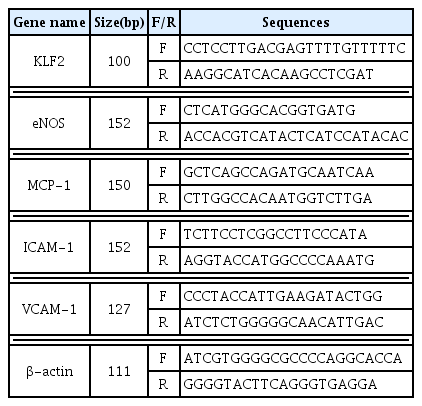
Table 1

Real-Time PCR Primer Sequences

Gene name Size(bp) F/R Sequences
KLF2 100 F CCTCCTTGACGAGTTTTGTTTTTC
R AAGGCATCACAAGCCTCGAT

eNOS 152 F CTCATGGGCACGGTGATG
R ACCACGTCATACTCATCCATACAC

MCP-1 150 F GCTCAGCCAGATGCAATCAA
R CTTGGCCACAATGGTCTTGA

ICAM-1 152 F TCTTCCTCGGCCTTCCCATA
R AGGTACCATGGCCCCAAATG

VCAM-1 127 F CCCTACCATTGAAGATACTGG
R ATCTCTGGGGGCAACATTGAC

β-actin 111 F ATCGTGGGGCGCCCCAGGCACCA
R GGGGTACTTCAGGGTGAGGA