The Effect of Geopungchunghyul-dan on Circumferential Strain of Carotid Artery: A Case Series

Article information

J Korean Med. 2018;39(1):86-94
Publication date (electronic) : 2018 March 31
doi : https://doi.org/10.13048/jkm.18009
1Department of Cardiology and Neurology, Kyung Hee University Korean Medicine Hospital
2Department of Cardiology and Neurology, College of Korean Medicine, Kyung Hee University
Correspondence to: 정민호 (Minho Jung), 경희대학교 한의과대학 순환신경내과학 교실, Tel: +82-2-958-9129, Fax: +82-2-958-9104, E-mail: tox37@naver.com
Received 2017 November 30; Revised 2018 March 5; Accepted 2018 March 2.

Abstract

Objectives

This study is aimed to examine the effect of Geopungchunghyul-dan on circumferential strain of carotid artery in outpatients of Kyung Hee University hospital of Korean Medicine.

Methods

Retrospective chart review is used from June 2016 to August 2017 for outpatients of Kyung Hee University hospital of Korean Medicine. 14 patients taking Geopungchunghyul-dan over 1 month were speculated. Data of circumferential strain and intima-media thickness was taken from carotid ultrasonography and processed by Wilcoxon signed-rank test.

Results

Geopungchunghyul-dan lowered circumferential strain of both carotid arteries after 1 month of administration (p<0.05). Intima-media thickness of both carotid arteries did not changed significantly.

Conclusions

Geopungchunghyul-dan may ameliorate arterial stiffness.

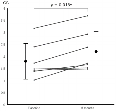
Fig. 1

Change of left circumferential strain for 1 month of treatment.

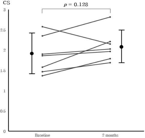
Fig. 2

Change of right circumferential strain for 1 month of treatment.

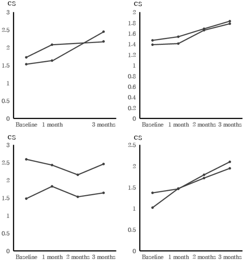
Fig. 3

Change of left circumferential strain for 2 months of treatment

Fig. 4

Change of right circumferential strain for 2 months of treatment

Fig. 5

Change of both circumferential strain for 3 months of treatment

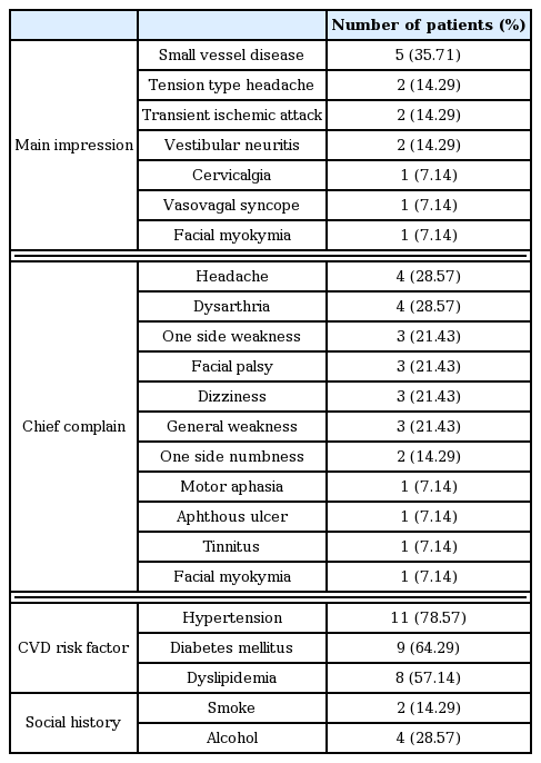
Clinical Characteristics of Patients. (n=14)

Mean Value and Standard Deviation of Circumferential Strain and Intima-Media Thickness of each Common Carotid Artery at Start(Baseline) and 1 Month of Treatment.

Rate of Change in Circumferential Strain for 1 Month of Treatment

References

1. Cohn JN, Duprez DA, Grandits GA. Arterial elasticity as part of a comprehensive assessment of cardiovascular risk and drug treatment. Hypertension 2005;46(1):217–220.
2. Boutouyrie P, Tropeano AI, Asmar R, Gautier I, Benetos A, Lacolley P. Aortic stiffness is an independent predictor of primary coronary events in hypertensive patients: a longitudinal study. Hypertension 2002;39(1):10–15.
3. Laurent S, Boutouyrie P, Asmar R, Gautier I, Laloux B, Guize L, et al. Aortic stiffness is an independent predictor of all-cause and cardiovascular mortality in hypertensive patients. Hypertension 2001;37(5):1236–1241.
4. Bjällmark A, Lind B, Peolsson M, Shahgaldi K, Brodin LA, Nowak J. Ultrasonographic strain imaging is superior to conventional non-invasive measures of vascular stiffness in the detection of age-dependent differences in the mechanical properties of the common carotid artery. Eur J Echocardiogr 2010;11:630–636.
5. Saji N, Toba K, Sakurai T. Cerebral small vessel disease and arterial stiffness: tsunami effect in the brain. Pulse 2016;3(3–4):182–189.
6. Jung WS, Kwon S, Cho SY, Park SU, Moon SK, Park JM, et al. The Effects of Chunghyul-Dan (A Korean Medicine Herbal Complex) on Cardiovascular and Cerebrovascular Diseases: A Narrative Review. Evid Based Complement Alternat Med 2016;2016:2601740.
7. Park HE, Cho GY, Kim HK, Kim YJ, Sohn DW. Validation of circumferential carotid artery strain as a screening tool for subclinical atherosclerosis. J Atheroscler Thromb 2012;19(4):349–56.
8. Catalano M, Lamberti-Castronuovo A, Catalano A, Filocamo D, Zimbalatti C. Two-dimensional speckle-tracking strain imaging in the assessment of mechanical properties of carotid arteries: feasibility and comparison with conventional markers of subclinical atherosclerosis. Eur J Echocardiogr 2011;Jul. 12(7):528–535.
9. Yuda S, Kaneko R, Muranaka A, Hashimoto A, Tsuchihashi K, Miura T, et al. Quantitative measurement of circumferential carotid arterial strain by two-dimensional speckle tracking imaging in healthy subjects. Echocardiography 2011;Sep. 28(8):899–906.
10. Yang EY, Dokainish H, Virani SS, Misra A, Pritchett AM, Lakkis N, et al. Segmental analysis of carotid arterial strain using speckle -tracking. J Am Soc Echocardiogr 2011;Nov. 24(11):1276–1284.
11. Saito M, Okayama H, Inoue K, Yoshii T, Hiasa G, Sumimoto T, et al. Carotid arterial circumferential strain by two-dimensional speckle tracking: a novel parameter of arterial elasticity. Hypertens Res 2012;Sep. 35(9):897–902.
12. Wierzbowska-Drabik K, Cygulska K, Cieślik-Guerra U, Uznańska-Loch B, Rechciński T, Trzos E, et al. Circumferential strain of carotid arteries does not differ between patients with advanced coronary artery disease and group without coronary stenoses. Adv Med Sci 2016;Sep. 61(2):203–206.
13. Boutouyrie P. Determinants of pulse wave velocity in healthy people and in the presence of cardiovascular risk factors: ‘establishing normal and reference values’ The Reference Values for Arterial Stiffness’ Collaboration. European Heart Journal 2010;31:2338–2350.
14. Park TH. Neuroprotective effect of Geopungchunghyul-dan on in vitro oxygen-glucose deprivation and in vivo permanent middle cerebral artery occlusion model[Dissertation] Seoul: Kyunghee Univ; 2015.
15. Park SU, Jung WS, Moon SK, Ko CN, Cho KH, Kim YS, et al. Chunghyul-dan(Qingxie-dan) improves arterial stiffness in patients with increased baPWV. Am J Chin Med 2006;34(4):553–563.
16. Min KD. The Effects of Cardiotonic Pills® on Common Carotid Artery Elasticity in Healthy Subjects [Dissertation] Seoul: Kyunghee Univ; 2016.
17. Park SU. Effect of Chunghyul-dan on Arterial stiffness: Clinical and Molecular Study [Dissertation] Seoul: Kyunghee Univ; 2005.
Article information Continued

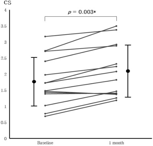
Fig. 1

Change of left circumferential strain for 1 month of treatment.

Fig. 2

Change of right circumferential strain for 1 month of treatment.

Fig. 3

Change of left circumferential strain for 2 months of treatment

Fig. 4

Change of right circumferential strain for 2 months of treatment

Fig. 5

Change of both circumferential strain for 3 months of treatment

Table 1

Clinical Characteristics of Patients. (n=14)

Number of patients (%)
Main impression Small vessel disease 5 (35.71)
Tension type headache 2 (14.29)
Transient ischemic attack 2 (14.29)
Vestibular neuritis 2 (14.29)
Cervicalgia 1 (7.14)
Vasovagal syncope 1 (7.14)
Facial myokymia 1 (7.14)

Chief complain Headache 4 (28.57)
Dysarthria 4 (28.57)
One side weakness 3 (21.43)
Facial palsy 3 (21.43)
Dizziness 3 (21.43)
General weakness 3 (21.43)
One side numbness 2 (14.29)
Motor aphasia 1 (7.14)
Aphthous ulcer 1 (7.14)
Tinnitus 1 (7.14)
Facial myokymia 1 (7.14)

CVD risk factor Hypertension 11 (78.57)
Diabetes mellitus 9 (64.29)
Dyslipidemia 8 (57.14)
Social history Smoke 2 (14.29)
Alcohol 4 (28.57)

Table 2

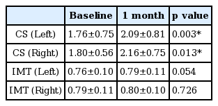
Mean Value and Standard Deviation of Circumferential Strain and Intima-Media Thickness of each Common Carotid Artery at Start(Baseline) and 1 Month of Treatment.

Baseline 1 month p value
CS (Left) 1.76±0.75 2.09±0.81 0.003*
CS (Right) 1.80±0.56 2.16±0.75 0.013*
IMT (Left) 0.76±0.10 0.79±0.11 0.054
IMT (Right) 0.79±0.11 0.80±0.10 0.726

CS, circumferential strain; IMT, intima-media thickness.

Wilcoxon signed-rank tests were used for comparison between start and 1 month of treatment.

*

Statistically significant at p<0.05

Table 3

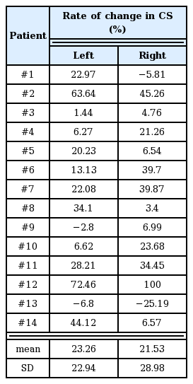
Rate of Change in Circumferential Strain for 1 Month of Treatment

Patient Rate of change in CS (%)

Left Right
#1 22.97 −5.81
#2 63.64 45.26
#3 1.44 4.76
#4 6.27 21.26
#5 20.23 6.54
#6 13.13 39.7
#7 22.08 39.87
#8 34.1 3.4
#9 −2.8 6.99
#10 6.62 23.68
#11 28.21 34.45
#12 72.46 100
#13 −6.8 −25.19
#14 44.12 6.57

mean 23.26 21.53
SD 22.94 28.98

CS, circumferential strain; SD, standard deviation