Non-operative Korean Medicine Treatment for Four Patients with Failed Back Surgery Syndrome after Spinal Fusion Surgery : A Retrospective Case Series
Article information
Abstract
Objectives
The purpose of this study was to report Four cases of Failed Back Surgery Syndrome (FBSS) patients after spinal fusion surgery who showed significant improvement in pain and function with Complex Korean medical treatment.
Methods
This study was a retrospective observational study. We reviewed medical records of Four patients with lumbar pain or radiating leg pain, who have received spinal fusion surgery in the past. All Four patients took complex treatments of Mokhuri Neck and Back hospital which involes Acupuncture, Pharmaco-acupuncture, Gangchuk herbal medicine, Chuna and Physical therapy during about four-week of admission treatment. Visual Analogue Scale (VAS), Oswestry disability index (ODI), Pain Free Walking Distance (PFWD) scores were assessed before and after treatments.
Results
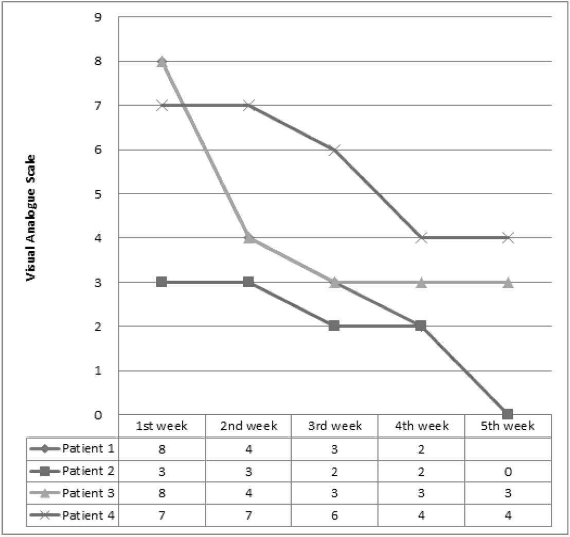
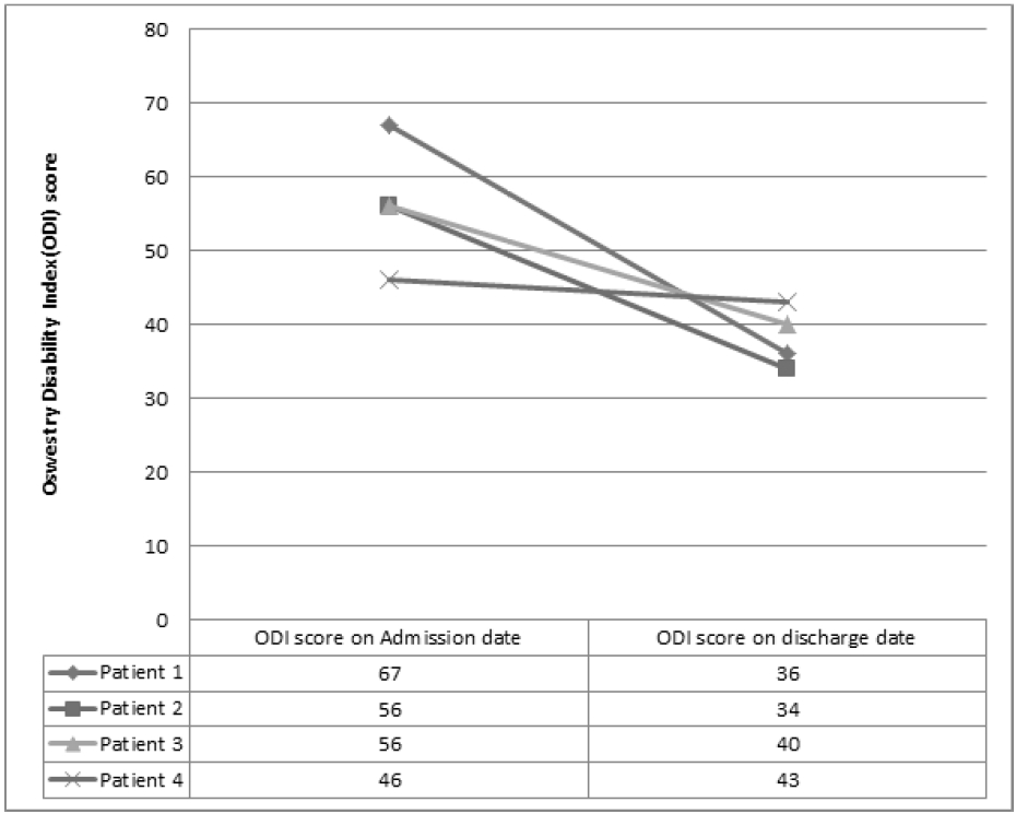
The average of hospitalization period was 28.5 days. Mean VAS scores decreased from 6.5 to 2.3, Oswestry Disability Index (ODI) scores decreased from 56.25 to 38.25 and Pain Free Walking Distance (PFWD) also improved from 10m to 166.6m.
Conclusion
This study implies that a combination of Korean medical treatments might be effective in relieving pain, and improving the functional status of FBSS patients. Further studies are needed to fully understand the mechanisms underlying the effects.
Introduction
Failed Back Surgery Syndrome (FBSS) is diagnosed in patients who have persistent back pain despite having undergone spinal surgery of any type, including discectomy, laminectomy, or fusion1). In a broad sense, FBSS is a term embracing a constellation of conditions that describes persistent or recurring Low Back Pain (LBP), with or without sciatica following one or more spinal surgeries2).
Failure of such surgeries is not rare. One study by Cauchoix et al evaluated the failure of surgery for 6%4). Another study by Javid et al reports a surgical failure rate as approximately 30%5).
Overall, The failure rate in spinal surgery is 15%, which suggests considerable potential risk of spinal surgery6). In addition, the success rate for secondary surgery after the initial failed surgery is also very low. Secondary surgery after failed spinal fusion showed a 50–60% fail rate and a third or fourth operation each showed to have a 50–60% and 70–80% fail rate. As the number of surgical procedures increased, the chance of having successful outcomes decreased3).
Considering high failure risk of spinal surgery, patients with FBSS are generally recommended to receive initial conservative treatment and consider surgical treatment only if conservative treatment shows to have no effect.
Among various types of conservative treatments, Korean medical treatments such as bee venom acupuncture and pharmaco-acupuncture have been reported to be a helpful intervention to the FBSS patients7–8). Generally, Herbal medicine, acupuncture, manual therapy (such as Chuna) and physiotherapy are used simultaneously for this condition in the current clinical practice of korea but there is no research on the benefit and harm of complex interventions available.
From these perspectives, we are reporting Four FBSS patients who expressed recurrent pain after fusion surgery due to Adjacent Segment Disease (ASD) and showed clinically significant improvements after about One month of complex korean medicine treatment at Mokhuri Neck & Back Hospital.
Methods
This study was an retrospective observational study based on the medical records of patients that received admission treatment at Mokhuri Neck & Back Hospital. All patients had received spinal fusion surgery of lumbar vertebra and showed recurrent lower back pain and leg pain. The data collected was from Jan. 2014. to Dec. 2015.
All patients that participated in the Research trial agreed on the usage of their medical records for research purposes The patients were told to notify medical officials if any adverse events were to occur.
1. Treatment
All patients received acupuncture treatment, oral administration of herbal medicine, pharmaco-acupuncture treatment, relaxative chuna therapy, and physical therapy for an average of Four weeks.
(1) Acupuncture treatment
Each patient received acupuncture treatment (2 sessions a day, 1 session on Sunday, total 13 sessions a week).
GV3, BL23, BL24, GB30 were treated in all patients, other acupuncture points were selected based on the symptoms of each patient by a Korean Medical Doctor.
0.25*40mm disposable stainless steel acupuncture needles (Dong Bang Co., Korea) were used for treatment. Acupuncture treatment time was about 15 minutes.
Everyday besides Sunday, patients received Two acupuncture sessions, One was given with electro-acupuncture and the other without electrical stimulation. On Sundays patients would only receive acupuncture without electrical stimulation
(2) Oral administration of herbal medicine
Each patient received oral administration of Gangchuk herbal decoction, a Herbal medicine three times a day, 30 minutes after each meal.
Gangchuk herbal decoction is composed of Geranium thunbergii which is known for its anti-nociceptive and anti-inflammatory effects, and Sorbus commixta which inhibits osteoclastogenesis and stimulates chondrogenesis along with many other herbs9) that in Korean medicine make the muscles and bones strong in order to reduce pain and strengthen the weakened muscle.
Other components of Gangchuk herbal decoction are Eucommiae cortex, Achyranthis radix, Cibotii rhizoma, Saposhnikoviae radix, geranii herba and Acanthopanacis cortex.
(3) Pharmaco-acupuncture treatment
Each patient received Hwangryunhaedok-tang pharmacoacupuncture injection treatment which consisted of Coptidis rhizoma, Scutellariae radix, Phellodendri cortex, and Gardeniae fructus extracts.
Hwangryunhaedok-tang pharmacoacupuncture solution (1–2 cc; Korean Pharmacoacupuncture Institute, Korea) was injected at the same site of acupuncture site subcutaneously.
Hwangryunhaedok-tang is a Korean medical prescription composed of Coptidis rhizoma, Scutellariae radix, Phellodendri cortex, and Gardeniae fructus extracts. Hwangryunhaedok-tang is distillated and used after many processes including cooling, filtration, pH regulation and high pressure sterilization.
Pharmacoacupuncture needles are 26-gauge insulin syringes and each patient received treatment once a day.
(4) Relaxative Chuna
Relaxative Chuna is manual Korean manipulation techniques which relaxes low back muscles including Latissimus dorsi, Rhomboid, Quadratus lumborum, Gluteus medius&minimus, paraspinal muscles, etc.
The patient laid prone on the ErgoStyleTM FX. -5820 table(Chattanooga Group. USA). Using the COX maneuver and manual manipulation, the lumbar spines were flexed and extended to 5~15 degrees 20 times a minute. This was to relieve the lumbar muscles and pelvic muscles. Each session lasted 15 minutes each and was held out five times a week.
(5) Physical therapy
Each patient received physical therapy 6 times a week (patients didn’t receive physical therapy on Sunday).
Physical therapy was consist of microwave therapy (5 minutes), Muscle low frequency therapy (10 minutes), Electromagnetic therapy (10 minutes) and Hot pack (10 minutes).
Physical therapy was applied on both Erector spinaes, Gluteus muscles, Iliolumbar ligaments and Sacroiliac ligaments.
2. Assessment
Visual Analogue Scale (VAS), Functional status of patient, Pain Free Walking Distance (PFWD) was used for assessment of patient’s condition during the admission period of each patient.
(1) VAS
VAS is a visual scale showing the degree of pain felt by the patient. A point is drawn on a 10cm long line where one end represents no pain and the other the worst pain possible to imagine. One is unable to use this score to compare between patients, however it is useful in recording the course of pain levels throughout a single patient. VAS scores were checked every week.
(2) Functional status of patient
Oswestry Disability Index (ODI) is an index to rate the degree of disability in the Lumbar region. It was first introduced in 1980 and is made up of 10 categories and 6 responses for each category. Each category is scored between 0 and 5. All ten scores answered are then added and divided by the total score possible (if all categories were answered, 50) then multiplied by 100. This result in a score ranging from 0~100. A higher score indicates a higher rate of disability.11) The ODI was completed two times during the treatment, Admission date and discharge date.
(3) PFWD
PFWD is the distance the patient is able to walk free of any back pain or radiating leg pain. The PFWD was measured every week in this study.
Case series
1. Patient 1 (Female, 77 years old)
-
- C/C
LBP
Both buttock/leg pain
-
- O/S
April 2014
-
- Past History
L4–5 Fusion Surgery (8 years ago)
-
- Family History
None specific
-
- Social History
None specific
- Present illness
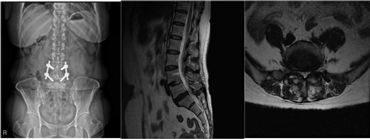
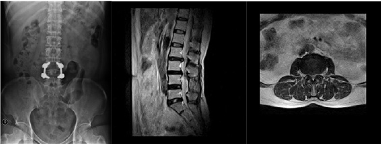
Received L4–L5 lumbar spinal fusion surgery 8 years ago (Figure 1). Received N-block treatment, April 2014 due to LBP and both buttocks/leg pain. Treatment was unsuccessful and persistent pain was experienced. HIVD in the L3 level was found in a Lumbar MRI test taken May 20 2014. Analgesics were taken afterwards but due to constant pain was admitted to Mokhuri Neck & Back Hospital July 1st, 2014. Patient was taking DM medication.
- Findings
-
- Diagnosis
Lumbar Disc protrusion of L2–3, L3–4
Post Fusion surgery state of L4–L5
-
- Hospitalization period
July 1, 2014 ~ July 26, 2014 (26 days)
- Progress(Table 1)
2. Patient 2 (Female, 77 years old)
-
- C/C
LBP
Both buttock/leg numbness (Lt>Rt)
-
- Past History
L4–5 Fusion Surgery (8 years ago)
-
- Family History
None specific
-
- Social History
None specific
- Present illness
Received lumbar spine Fusion surgery 8 years ago. Recurrent pain became worse in March 2014, March 22, patient took a Lumbar MRI and was recommended surgery but the patient refused. Instead, underwent neuroplasty, failed to relieve of pain and started Per Oral (PO) medication. Visited Mokhuri Neck & Back Hospital July 24th 2014. Admitted July 28th 2014. Patient was on HTN medication.
- Findings
-
- Diagnosis
Post Fusion Surgery state of L4–5
Spondylolisthesis of L4 on L5, Grade 1.
Spinal stenosis of L4–5
Broad-based disc protrusion of L5-S1
-
- Hospitalization period
July 28, 2014 ~ August 23, 2014 (27 days)
- Progress(Table 2)
3. Patient 3 (Female, 55 years old)
-
- C/C
Rt LBP
Rt leg radiating pain&numbness
-
- Past History
L4–5 Fusion Surgery (6 years ago)
-
- Family History
None specific
-
- Social History
None specific
- Present illness
Received Lumbar fusion surgery 6 years ago. Attended to a local OS whenever pain was felt and received Physical therapy and PO medication. Pain intensified December of 2014 and visited Mokhuri Neck & Back Hospital. Unable to extend L-spine and walked with a limp due to pain. Unable to sit still for 5 minute due to pain.
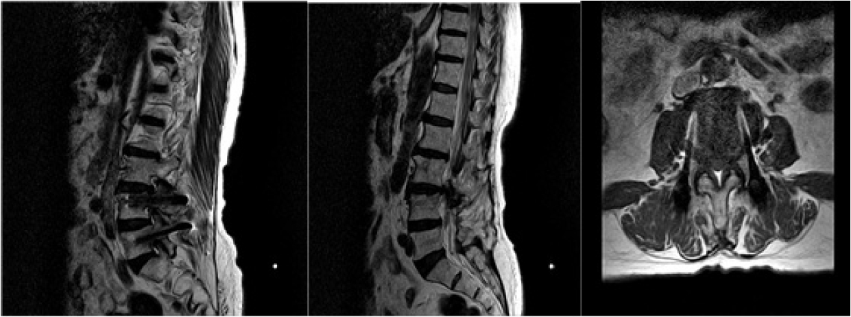
- Findings(Figure 3)
-
- Diagnosis
a. L2–3 - Right central disc protrusion with annular tear
b. L3–4 - Diffuse bulging disk
- Spondylolisthesis of L3 on 4, grade 1
-
- Facet joint arthrosis at both sides --> Narrowed spinal canal, suggested
c. L4–5 - Spinal fusion with pedicle screw state
d. L5-S1 - Central disc protrusion
e. Degenerative disc change at L2–3 to L5-S1
-
- Hospitalization period
February 24, 2014 ~ March 22, 2014 (28 days)
- Progress(Table 3)
4. Patient 4 (Female, 75 years old)
-
- C/C
LBP
Both buttock pain
Both leg radiating pain
-
- Past History
Fusion surgery (3 years ago)
-
- Family History
None specific
-
- Social History
None specific
- Present illness
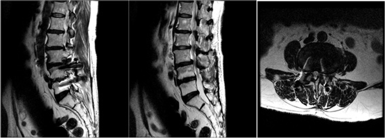
Symptoms first appeared in 2009, after L-spine MRI test patient was diagnosed with Lumbar stenosis. Received Fusion surgery in L4–5 levels in 2012. Patient experienced continuous pain and in Feb. 2014 took another L-spine MRI exam and was diagnosed with ASD and spondylolisthesis grade 2. Received nerve block numerous times but failed to be relieved of pain and visited Mokhuri Neck & Back Hospital.
Patient was taking HTN and DM medication
Both leg pain and tension was felt on first step of walking and had trouble standing still.
- Findings(Figure 4)
-
- Diagnosis
L1–2 - Rt subarticular disc protrusion
L2–3 - Lt subarticular disc protrusion
L3–4 - Spondylolisthesis L3 on L4, Grade 1, spinal stenosis
L4–5 - modic type 2 change of end plate, post fusion surgery state.
-
- Hospitalization period
September 7, 2015 ~ October 9, 2015(33 days)
- Progress(Table 4)
Results
The 4 patients mentioned above all went through Spinal Fusion surgery. After surgical treatment, Disc herniation or spondylolisthesis, recurrent LBP and leg pain occurred. All 4 patients had pain or gait disturbance that restricted the patients from everyday activities. All patients received admission treatment and the average hospitalization period was 28.5 days. Average VAS scores were 6.5 before treatment and reduced to 2.3 after treatment (Figure 5) ODI scores were average 56.25 before admission treatment and showed improved to 38.25 after treatment (Figure 6). The PFWD also showed improvement from an average of 10m before treatment to an average of 166.6m (Figure 7). Minor adverse events such as slight bleeding or bruises occurred after acupuncture treatment. But no severe adverse events such as a drop in functional status or intensified pain were reported
Discussion
The results of this Retrospective Case Series shows that with about 1 month (average 28.5 days) of Complex Korean Medical Treatment.
The mechanism of this complex intervention is not established currently. However, each intervention has therapeutic effect and all individual effect might be summed up for relieving symptoms of FBSS. The effects of acupuncture on LBP can be found in multiple previous studies and its clinical efficiency is acknowledged among many practitioners.12–15). Brinkhaus B, et al. reported that the group which had received acupuncture had a significantly higher analgesic effect compared to the non-acupuncture group13). Zhao. Z. Q. reported that manual acupuncture stimulated Aβ-fibers as well as a part of Aδ-fibers which results as an analgesic effect15). These analgesic natures of acupuncture are seemed to be the reasons for reducing pain in FBSS patients. Pharmacoacupuncture treatment used in this study was initially designed to combine the effects of acupuncture and herbal medicine. Hwangryunhaedok-tang pharmacoacupuncture solution was used in this study. Hwangryunhaedok-tang pharmacoacupuncture is used in many painful diseases and there are reports of its anti-inflammatory actions.16) The underlying reason for pain in FBSS can originate from many reasons. Besides from problems involving the surgical procedure itself or misdiagnosis, Common reasons include inflammations in the arachnoid or chronic inflammations related to surgery which interfere with the mobility of the nerve root itself causing pain17)18), and epidural fibrosis19). Which can explain why Hwangryunhaedok-tang pharmacoacupuncture is effective. The clinical evidence of Chuna treatment has been suggested: 19 papers about Chuna treatment were published in the peer-reviewed journals after 2010. In acute LBP patients a maximum of 4 weeks of treatment and in chronic LBP patients a range of 8~12 weeks of treatment showed to be effective.20) Relaxative Chuna used in this study relieves stress in the muscles of the lower back including the Latissimus dorsi, Rhomboids, Quadratus lumborum, Gluteus medius & minimus, and the paraspinal muscles. By relieving stress in the lower back is thought to help reduce pain.
Ganchuk-tang, a herbal medicine for this condition include several medicinal herbs that in Korean medicine make the muscles and bones strong in order to reduce pain and strengthen the weakened muscle and each of them has therapeutic effect on FBSS patients
Although the results of this study are positive, there are several limitations. First of all we were only able to analyze data during the average 4 weeks of admission treatment. Only the short-term effects of the treatment were shown and data after discharge was scarce and so it was unable to analyze the long-term effects. Second there is only small number of patient cases so we can only provide a limited result. Third, admission treatment was a combination of treatments and it is impossible to fully understand the effects of each treatment involved. It seems necessary to design additional studies for overcoming this limitation of this study.
There are many studies reporting the effects of Complex Korean Medical Treatment on Lumbar disc herniation or Spinal stenosis. However there were no studies regarding the effects of Complex Korean Medical Treatment in recurrent pain due to FBSS after Fusion surgery with ASD. In this study, Four cases of patients showed improvement in pain, PFWD, and functional status after complex Korean Medicine treatment. In the future, rigorous randomized clinical trials should be designed to verify the clinical efficacy of Complex Korean Medical treatment.
