A Research of characteristics of left/right pulse wave and blood vessel using Korean medicine pulse diagnosis
Article information
Abstract
Objectives:
The pulse diagnosis to identify the symptoms has been considered important in Korean medicine. The position and character of disease would be confirmed by pulse diagnosis of left and right radial artery. This paper is to analyze the characteristics and differences of left and right blood vessels.
Methods:
In this study, left and right radial artery and dorsalis pedis artery was measured and analyzed by using condenser typed pulse analyzer. Commercially available pulse analyzer was used to measure the radial artery. The pulse wave was measured in 20 laboratory healthy men and women. The blood vessel aging degree and index of augmentation of blood vessel was obtained from the measured pulse wave graph and the characteristics and differences of the left and right blood vessel was analyzed.
Results:
The significant difference of pulse transit time between the right handed and non-right handed was not found. The correlation of radial artery and dorsalis pedis artery had no significant difference. By obtaining the blood vessel aging index (AGI) and augmentation index (AI) of blood vessel at the left and right radial artery, the significant difference between right handed and non-right handed was not found.
Conclusions:
The result of this study would help to explain the characteristic of blood vessel with respect to the left and right handed. We suggest that research of pulse wave of the left and right blood vessel using pulse analyzer should be needed in further study.
Measurement procedure of the experiment
Simultaneous measurement of two arterial pulse signals at gwan region of both right and left hand by using pulse diagnosis of condenser microphone.
(a) The parameter of the pressure pulse wave graph (b) The velocity pulse wave (primary derivative) (c) The acceleration pulse wave (second derivative)
The various acceleration pluse wave types. Younger people are toward (a) type and older people are toward (g) type.
Pulse transit time at left/right radial artery and dorsalis pedis artery
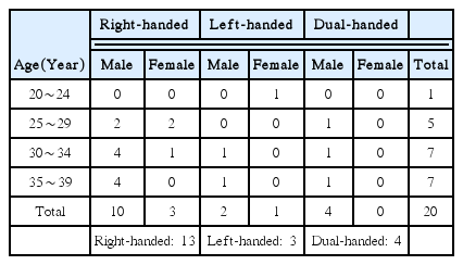
Age and Gender Distribution of the Experiment
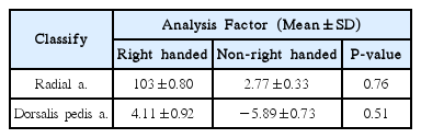
Comparision of Right Handed and Non-Right Handed of the PTT and Radial Artery and Dorsalis Pedis Artery
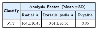
Comparision of PTT between Radial Artery and Dorsalis Pedis Artery
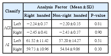
Comparision of Right Handed and Non-Right Handed of the AGI and AI
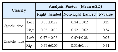
Comparision of Right Handed and Non-Right Handed of Systole and Diastole Time